Page 1 :
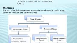
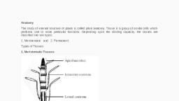
ANATOMY OF FLOWERING PLANTS, ✓, ✓, ✓, ✓, ✓, ✓, , ✓, ✓, , ✓, ✓, , Tiaaue, A tissue is group of cells having common origin and performing a common function ., There are two types of Tissues in plants, a) Permanent tissue and b) meristematic tissue, MERISTAMATIC TISSUES, Growth in plants is largely restricted to specialised regions of active cell division called, meriatema., Based on its position in the plant body, the meristem is divided into three types -apical, m enstem, ·, ·, al arv menstem, ·, and lateral meristem ., . mterc, Lateral med.atem, lntercalary meriatem, Apical meriatem, , ., , ., , ., , • Occurs at the tips of, roots and shoots, • Primary meristem, • Increase the length of, plant, , ·Occurs between mature tissue, at nodal region, • Primary meristem, • Help the grasses to regenerate, parts removed by grazing, herbivores., , · Present along the sides of, the plant body, • Secondary meris tern, • Appears later stage of, growth and responsible for, increase in thickness ., , Both apical meristems and intercalary meristems are primary meristems because they appear, early in life of a plant and contribute to the formation of the primary plant body., Axillary bud : The buds which are present in the axils of leaves and are responsible for forming, branches or flowers., COMPLEX T ISSUES, Complex tissue consists of different types of cells but perform same function., Xylem and phloem constitute the complex tissues in plants and work together as a unit., XYLEM, , ✓, , ✓, , Xylem functions as tissue for transport of, water and minerals from roots to the stem- and leaves., Xylem is composed of four different types of, elements, namely, tracheida, v essel.a, xylem, fibre• and xylem parenchyma., , ✓, ✓, , ✓, ✓, , TradW!ld, , ✓, ✓, , ✓, ✓, ✓, , ✓, , ✓, ✓, , Protoxylem & metaxylem, Primary xylem is of two types - protoxylem, and metaxylem., The first formed primary xylem elements are, called protoxylem and the later formed, primary xylem is called metaxylem, , ✓, , ✓, , Tracheids are elongated or tube like cells with, thick and lignified walls and tapering ends., Vessel is a long cylindrical tube-like structure, made up of many cells called vessel members ., Gymnosperms lack vessels in their xylem ., Vessel members are inter-connected by, perforations in their common walls., In flowering plants tracheids and vessels are, the main water transporting elements., Xylem fibers are dead cells with lignified cell, wall and central lumen., Xylem parenchyma are living and thin-walled, cells, and their walls are made up of cellulose., Radial conduction of water takes place by, specialise ray parenchyma cell.a., In stems, the protoxylem lies towards the, centre (pith) and the metaxylem lies towards, the periphery of the organ, this type of, primary xylem is called endarch., In roots, the protoxylem lies towards, periphery and metaxylem lies towards the
Page 2 :
✓, , ✓, , centre, such arrangement of primary xylem is, PHLOEM, Phloem transports food materials, usually, from leaves t o other parts of the plant., Phloem in angiosperms is composed of sieve, tube elements, companion cells, phloem, parenchyma, and phloem fibres., , J, , I, , /, , I, , ., , i:, , ~ ,, , ', , I, I, , , ' ,!, I, , :•urw, pore, -~, , tu b<', , ✓, , ✓, ✓, , ✓, , ✓, , ✓, , ✓, , ✓, , ✓, ✓, ✓, , ✓, ✓, , ✓, , f'k-1:Mol, , pwud»,1:Qil, , .., , Compan.lOII, , ,.,., , l, , o,,, ✓, , called exarch, , Instead of sieve tubes and companion cells, Gymnosperms ha ve albu.minoll8 cell■ and, aleve cell■ ., Sieve tube elements are also long, tu be-like, s tructures , arranged longitudinally and are, associated with the companion cella., Their end walls are perforated in a sieve-like, manner to form the sieve plates., A mature sieve element possesses a peripheral, , ✓, ✓, ✓, , ✓, ✓, , ✓, , cytoplasm and a large vacuole but lacks a, nucleus., The functions of sieve tu bes are controlled by, the nucleus of companion cell■•, The companion cells are specialised, parenchymatous cells, which are closely, associated with sieve tu be elements., Phloem parenchyma is made up of elongated,, cylindrical cells which have dense cytoplasm, and nucleu s., Phloem parenchyma is a bsent in most of the, mon ocotyledons., Phloem fibres (bast fibres) are m ade up of, sclerenchymatous cells., These are generally absent in the primary, phloem but are found in the secondary, phloem., Phloem fibres of jute, flax and hemp are used, commercially., The first formed primary phloem consists of, narrow sieve tu bes and is referred to as proto, phloem., Later formed phloem has bigger sieve tu bes, and is referred to as meta phloem., , THE TISSUE SYSTEM, On the basis of their location and function all the tissues of a plant can be classified in to three, tissue systems., EPIDERMAL TISSUE SYSTEM, trans piration and gaseous exchange., It forms the outermost covering of whole, ✓ Each s toma is composed of two bean, plant body, which consis ts of e pidermal, shaped cells known as pa.rd cell• which, cells, stomata, epidermal appendages, enclose stomata! pore., (trichomes and hairs) ., ✓ Epidermal cells adjacent to the guard cells, Epidermis forms outermost protecting layer, become specialised in their shape and size, made up of i:s single layered paren chyma, and are known as aubaidiary cella., cells., ✓ The s toma ta! a perture, guard c,e lls and the, The outside of the epidermis is often covered, surrounding subsidiary cells are together, with a waxy thick layer called the cuti.cle, called ■tomatal apparatll8., which prevents the loss of water ., ✓ The root hairs are unicellular elongation of, Cuticle is absent in roots., e pidermal cells and help in absorption of, Stomata are structures present in the, water and mineral nutrients., epidermis of leaves., ✓ Trich.o me• arc multicellular h airs present, Stomata regulate the process of, on the stem., VASCULAR TISSUE SYSTEM, The vascular system consists of complex, tissues, xylem and phloem that together
Page 3 :
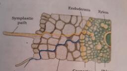
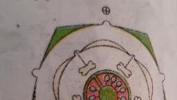
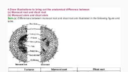
✓, ✓, , ✓, , ✓, , form vaacular bundle,., the arrangement of xylem and phloem are, different in roots and stem., In roots the xylem and phloem seen in, different radii. Such vascular bundles are, called radial vascular bundles .(Roots), In stem the xylem and phloem are arranged, in the same radius. Such vascular bundles, , ✓, ✓, , ✓, , ANATOMY OF DICOT ROOT, ✓, Jr- - Root hntr, , E:pllknnts, , ✓, , Coru,x, , ✓, t::ndoc:1.-nm,o, l'tt--, , l'M"lt')~l.-, , n,--, , f'roloxvkm, , w- -llkl,Ul}'lm1, , ✓, , ✓, , Olcot Root, ✓, ✓, , are called conjoint(stem and leaves)., Conjoint vascular bundles may be open or, closed, In open vascular bundle cambium ( a, meristem) is seen in between xylem and, phloem as in dicot stem, In closed vascular bundle cambium is, absent as in monocot stem., , The outermost layer of dicot root is, epiblema containing unicellular root hairs., The cortex consists of several layers of thinwalled parenchyma cells., , ✓, , The innermost layer of cortex is called, endodermis,, Endodermal cell wall has thick deposition of, waxy material suberin,which is impermeable, to water,it is called caaparian strip., Inner to endodermis lies a few layers of, thick-walled parenchyomatous cells referred, to as, pericycle., Xylem and phloem groups are limited in, number .Xylem elements are polygonal in, outline., The parenchymatous cells which lie between, the xylem and the phloem are called, conjuctive tissue., All tissues on the inner side of the, endodermis such as pericycle, vascular, bundles and pith constitute the stele., , ANATOMY OF MONOCOT ROOT, , ✓, ✓, ✓, , ✓, , The anatomy of the monocot root is similar, to the dicot root in many respects., It has epidermis, cortex, endodermis ,, pericycle, vascular bundles and pith., Xylem and phloem groups are more in, number than dicot root., Pith is large and well developed., , Monoc:ot Root, ✓, , Dicot Root, 1. Cortex is comparatively narrow, , ✓, , Monocot Root, l. Cortex is very wide.
Page 4 :
✓, , 2. Endodermis is less thic kened casparian, stripes are more promine nt, 3. The xylem and phloem bundles varies, from 2 to 6, ~ - Pith is reduced or sometimes absent, 5 Secon dary growth takes place with the, h e h elp of cambium., ANATOMY OF DICOT STEM, J;plo!;:r.-. ,,, , "'", , .., , -·---, , ;· t:pJOttu•, , ~ -, , 1 ··">'$l"d-'""', ' \tN'Uli '\yt'hl, , - t:CuJCll!it.TCIU,, , ✓, ✓, , ✓, , ✓, , ✓, , ✓, , ✓, ✓, , ✓, ✓, , Q . Casparian strips are visible only in young, , !Roots, O. Xylem and phloem are more than 6., (polyarc h) ., 14. Well developed and large pith is presen t., 5 . Secondary growth is a bs ent., , parenchymatous cells with intercellular, spaces., ✓ Endodermfa: The innermost layer of the, cortex rich in starch grains and hence called, Starch sheath, ✓ Pericycle is presen t on the inner side of the, endodermis and above the phloem., ✓ Vascular bundles limited in number and, arranged in a ring form., ✓ Vascular bundles are conjoint and open., ✓, Paren-chymatous tissue seen in between, vascular bundles constitute medullary, , Epidermis forms the outermost protective, layer of the stem., rays., The multiple layers of cells arranged in, ✓ A large number of rounded, parenbetween epidermis and pericycle constitu te, chymatous cells occupy the central portion, the cortex., of the stem constitute the pith., It has three sub-zones, ✓ Conjoint vascular bundles may be open or, Hypodennis.: Layers of collenchymatous, closed, cells just below the epidermis, which providemechanical strength to the young stem., - ✓ In ope n vascular bundle cambium ( a, meristem) is seen in between xylem and, Cortical layers: Layers of round thin walled, p hloem as in dicot stem., ANATOMY OF MONOCOT STEM, The hypodennis is made up of, sclerenchyma., Vascular bundles are conjoint and closed., Vascular bundles are numerous and, scattered in the ground tissue, Each vascular bundle is surrounded by a, sclerenchymatous bundle sheath., Phloem parenchyma is absent in monocot, stem and they have water-containing, cavities within the vascular bundles., , ••, •••, , Dicot Stem, 1. The ground tissue is differentiated into, ortex, endodermis, pericycle and p ith ., . The vascular bundles are arranged in a, g, , onocot Stem, 1. The ground tissue is made u p of similar, ells . (Homogeneous), . The vascular bundles are scattered in the, ound tissue.
Page 5 :
. Vascular bundles a re open, without, undle sheath., . The stem shows secondary growth., F, , . Vascular bundles are closed,and, s urrounded, y sclerenchymatous bundle sheath ., . Secondary growth is a bsent., , I ENTRAL (DICOT) LEAF, , ✓ The leaf lamina of a dorsiventral leaf has 3 parts: epidermis, meaophyll and vascular system., ✓ The upper ep idermis is called adaxial epidermis and lower one is called abaxial epidermis., ✓ More numbe r of stomata are present on the abaxial epidermis., , ✓ There are two types of cells in the mesophyll,upper layer called palisa de parenchyma and lower, , spongy parenchyma., ✓ There are numerous large spaces and air cavities between the cells of spongy parenchyma., ✓, , Vascular bundles can be seen in the midrib and veins., ✓ Vascular bu n dles are surrounded by a layer of t hick-walled bundle sheath cella.