Page 1 :
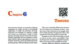
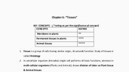
C hapter 6, TISSUES, From the last chapter, we recall that all living, organisms are made of cells. In unicellular, organisms, a single cell performs all basic, functions. For example, in Amoeba, a single, cell carries out movement, intake of food and, respiratory gases, respiration and excretion., But in multi-cellular organisms there are, millions of cells. Most of these cells are, specialised to carry out a few functions. Each, specialised function is taken up by a different, group of cells. Since these cells carry out only, a particular function, they do it very, efficiently. In human beings, muscle cells, contract and relax to cause movement, nerve, cells carry messages, blood flows to transport, oxygen, food, hormones and waste material, and so on. In plants, vascular tissues conduct, food and water from one part of the plant to, other parts. So, multi-cellular organisms, show division of labour. Cells specialising in, one function are often grouped together in, the body. This means that a particular, function is carried out by a cluster of cells at, a definite place in the body. This cluster of, cells, called a tissue, is arranged and designed, so as to give the highest possible efficiency of, function. Blood, phloem and muscle are all, examples of tissues., A group of cells that are similar in, structure and/or work together to achieve a, particular function forms a tissue., , 6.1 Are Plants and Animals Made, of Same Types of Tissues?, Let us compare their structure and functions., Do plants and animals have the same, structure? Do they both perform similar, functions?, , There are noticeable differences between, the two. Plants are stationary or fixed – they, don’t move. Most of the tissues they have are, supportive, which provides them with, structural strength. Most of these tissues are, dead, since dead cells can provide mechanical, strength as easily as live ones, and need less, maintenance., Animals on the other hand move around, in search of food, mates and shelter. They, consume more energy as compared to plants., Most of the tissues they contain are living., Another difference between animals and, plants is in the pattern of growth. The growth, in plants is limited to certain regions, while, this is not so in animals. There are some, tissues in plants that divide throughout their, life. These tissues are localised in certain, regions. Based on the dividing capacity of the, tissues, various plant tissues can be classified, as growing or meristematic tissue and, permanent tissue. Cell growth in animals is, more unifor m. So, there is no such, demarcation of dividing and non-dividing, regions in animals., The structural organisation of organs and, organ systems is far more specialised and, localised in complex animals than even in very, complex plants. This fundamental difference, reflects the different modes of life pursued, by these two major groups of organisms,, particularly in their different feeding methods., Also, they are differently adapted for a, sedentary existence on one hand (plants) and, active locomotion on the other (animals),, contributing to this difference in organ system, design., It is with reference to these complex, animal and plant bodies that we will now talk, about the concept of tissues in some detail.
Page 2 :
Q, , uestions, , •, , 1. What is a tissue?, 2. What is the utility of tissues in, multi-cellular organisms?, , 6.2 Plant Tissues, , From the above observations, answer, the following questions:, 1. Which of the two onions has longer, roots? Why?, 2. Do the roots continue growing even, after we have removed their tips?, 3. Why would the tips stop growing in, jar 2 after we cut them?, , The growth of plants occurs only in certain, specific regions. This is because the dividing, tissue, also known as meristematic tissue, is, located only at these points. Depending on, the region where they are present,, meristematic tissues are classified as apical,, lateral and intercalary (Fig. 6.2). New cells, produced by meristem are initially like those, of meristem itself, but as they grow and, mature, their characteristics slowly change, and they become dif ferentiated as, components of other tissues., , 6.2.1 MERISTEMATIC TISSUE, , Apical meristem, , Jar 1, , Jar 2, Intercalary meristem, , Fig. 6.1: Growth of roots in onion bulbs, , Activity ______________ 6.1, •, •, , •, •, •, , Take two glass jars and fill them with, water., Now, take two onion bulbs and place, one on each jar, as shown in, Fig. 6.1., Observe the growth of roots in both the, bulbs for a few days., Measure the length of roots on day 1,, 2 and 3., On day 4, cut the root tips of the onion, bulb in jar 2 by about 1 cm. After this,, observe the growth of roots in both the, jars and measure their lengths each, day for five more days and record the, observations in tables, like the table, below:, , Length, Jar 1, Jar 2, TISSUES, , Day 1, , Day 2, , Day 3, , Day 4 Day 5, , Lateral meristem, , Fig. 6.2: Location of meristematic tissue in plant body, , Apical meristem is present at the growing, tips of stems and roots and increases the, length of the stem and the root. The girth of, the stem or root increases due to lateral, meristem (cambium). Intercalary meristem is, the meristem at the base of the leaves or, internodes (on either side of the node), on twigs., 69
Page 3 :
As the cells of this tissue are very active,, they have dense cytoplasm, thin cellulose, walls and prominent nuclei. They lack, vacuoles. Can we think why they would lack, vacuoles? (You might want to refer to the, functions of vacuoles in the chapter on cells.), , •, , 6.2.2 PERMANENT TISSUE, , •, , What happens to the cells for med by, meristematic tissue? They take up a specific, role and lose the ability to divide. As a result,, they form a permanent tissue. This process, of taking up a permanent shape, size, and a, function is called differentiation. Cells of, meristematic tissue differentiate to form, different types of permanent tissue., , Now, answer the following on the basis, of your observation:, 1. Are all cells similar in structure?, 2. How many types of cells can, be seen?, 3. Can we think of reasons why there, would be so many types of cells?, We can also try to cut sections of plant, roots. We can even try cutting sections, of root and stem of different plants., , 6.2.2 (i) SIMPLE PERMANENT TISSUE, A few layers of cells form the basic packing, tissue. This tissue is parenchyma, a type of, permanent tissue. It consists of relatively, unspecialised cells with thin cell walls. They, are live cells. They are usually loosely packed,, , Trichome, Mucilaginous canal, Cuticle, Epidermis, Hypodermis, Cortex, Endodermis, Pericycle, Phloem, Cambium, Metaxylem, Protoxylem, Medullary ray, Xylem, Vascular bundle, Pith, , Fig. 6.3: Section of a stem, , Activity ______________ 6.2, •, , Take a plant stem and with the help, of your teacher cut into very thin slices, or sections., Now, stain the slices with safranin., Place one neatly cut section on a slide,, and put a drop of glycerine., Cover with a cover-slip and observe, under a microscope. Observe the, various types of cells and their, arrangement. Compare it with Fig. 6.3., , •, , •, , 70, , so that large spaces between cells, (intercellular spaces) are found in this tissue, [Fig. 6.4 a(i)]. This tissue provides support to, plants and also stores food. In some, situations, it contains chlorophyll and, performs photosynthesis, and then it is called, chlorenchyma. In aquatic plants, large air, cavities are present in parenchyma to give, buoyancy to the plants to help them float., Such a parenchyma type is called, aerenchyma. The parenchyma of stems and, roots also stores nutrients and water., SCIENCE
Page 4 :
The flexibility in plants is due to another, permanent tissue, collenchyma. It allows, easy bending in various parts of a plant (leaf,, stem) without breaking. It also provides, mechanical support to plants. We can find, , this tissue in leaf stalks below the epidermis., The cells of this tissue are living, elongated, and irr egularly thickened at the, corners. There is very little intercellular, space (Fig. 6.4 b)., , Intercellular spaces, , Wall thickenings, Nucleus, Vacuole, Cell wall, , a (i), , b (i), , Cytoplasm, Nucleus, End wall, Primary cell wall, (thickened at corners), , Middle lamella, , Chloroplast, Chloroplast, Nucleus, Vacuole, , Vacuole, , Cytoplasm, Intercellular space, , Intercellular space, , Primary cell wall, , a (ii), , b (ii), , Narrow lumen, Lignified, thick wall, , Simple, pit pair, , c (i), , c (ii), , Fig. 6.4: Various types of simple tissues: (a) Parenchyma (i) transverse section, (ii) longitudinal section;, (b) Collenchyma (i) transverse section, (ii) longitudinal section; (c) Sclerenchyma (i) transverse section,, (ii) longitudinal section., TISSUES, , 71
Page 5 :
Yet another type of permanent tissue is, sclerenchyma. It is the tissue which makes, the plant hard and stiff. We have seen the, husk of a coconut. It is made of, sclerenchymatous tissue. The cells of this, tissue are dead. They are long and narrow as, the walls are thickened due to lignin (a, chemical substance which acts as cement and, hardens them). Often these walls are so thick, that there is no internal space inside the cell, (Fig. 6.4 c). This tissue is present in stems,, around vascular bundles, in the veins of, leaves and in the hard covering of seeds and, nuts. It provides strength to the plant parts., , Activity ______________ 6.3, •, •, , Take a freshly plucked leaf of Rheo., Stretch and break it by applying, pressure., While breaking it, keep it stretched, gently so that some peel or skin, projects out from the cut., Remove this peel and put it in a petri, dish filled with water., Add a few drops of safranin., Wait for a couple of minutes and then, transfer it onto a slide. Gently place a, cover slip over it., Observe under microscope., , •, , •, •, •, , •, , Guard, cells, Stomata, Epidermal, cell, (a), , Guard, cell, (b), , Fig. 6.5: Guard cells and epidermal cells: (a) lateral, view, (b) surface view, , What you observe is the outermost layer, of cells, called epidermis. The epidermis is, usually made of a single layer of cells. In some, plants living in very dry habitats, the, 72, , epidermis may be thicker since protection, against water loss is critical. The entire, surface of a plant has this outer covering of, epidermis. It protects all the parts of the plant., Epidermal cells on the aerial parts of the plant, often secrete a waxy, water-resistant layer on, their outer surface. This aids in protection, against loss of water, mechanical injury and, invasion by parasitic fungi. Since it has a, protective role to play, cells of epidermal, tissue form a continuous layer without, intercellular spaces. Most epidermal cells are, relatively flat. Often their outer and side walls, are thicker than the inner wall., We can observe small pores here and there, in the epidermis of the leaf. These pores are, called stomata (Fig. 6.5). Stomata are, enclosed by two kidney-shaped cells called, guard cells. They are necessary for, exchanging gases with the atmosphere., Transpiration (loss of water in the form of, water vapour) also takes place through, stomata., Think about which gas may be required, for photosynthesis., Find out the role of transpiration in plants., Epidermal cells of the roots, whose, function is water absorption, commonly bear, long hair-like parts that greatly increase the, total absorptive surface area., In some plants like desert plants,, epidermis has a thick waxy coating of cutin, (chemical substance with waterproof quality), on its outer surface. Can we think of a reason, for this?, Is the outer layer of a branch of a tree, different from the outer layer of a young stem?, As plants grow older, the outer protective, tissue undergoes certain changes. A strip of, secondary meristem replaces the epidermis, of the stem. Cells on the outside are cut off, from this layer. This forms the several-layer, thick cork or the bark of the tree. Cells of, cork are dead and compactly arranged, without intercellular spaces (Fig. 6.6). They, also have a chemical called suberin in their, walls that makes them impervious to gases, and water., SCIENCE
Page 6 :
Cork cells, , Ruptured epidermis, , parts of the plant. Except for phloem fibres,, phloem cells are living cells., , Fig. 6.6: Protective tissue, , 6.2.2 (ii) COMPLEX PERMANENT TISSUE, The different types of tissues we have, discussed until now are all made of one type, of cells, which look like each other. Such, tissues are called simple permanent tissue., Yet another type of permanent tissue is, complex tissue. Complex tissues are made of, more than one type of cells. All these cells, coordinate to perform a common function., Xylem and phloem are examples of such, complex tissues. They are both conducting, tissues and constitute a vascular bundle., Vascular or conductive tissue is a distinctive, feature of the complex plants, one that has, made possible their survival in the terrestrial, environment. In Fig. 6.3 showing a section of, stem, can you see different types of cells in, the vascular bundle?, Xylem consists of tracheids, vessels,, xylem parenchyma (Fig. 6.7 a,b,c) and xylem, fibres. The cells have thick walls, and many, of them are dead cells. Tracheids and vessels, are tubular structures. This allows them to, transport water and minerals vertically. The, parenchyma stores food and helps in the, sideways conduction of water. Fibres are, mainly supportive in function., Phloem is made up of four types of, elements: sieve tubes, companion cells,, phloem fibres and the phloem parenchyma, [Fig. 6.7 (d)]. Sieve tubes are tubular cells with, perforated walls. Phloem is unlike xylem in, that materials can move in both directions in, it. Phloem transports food from leaves to other, TISSUES, , Xylem, , Phloem, , Nucleus, , Pit, , Pits, Cytoplasm, , (a) Tracheid, , (b) Vessel, , (c) Xylem parenchyma, , Sieve plate, Sieve tube, , Phloem, parenchyma, Companion cell, , (d) Section of phloem, , Fig. 6.7: Types of complex tissue, 73
Page 7 :
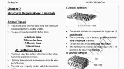
Q, , uestions, 1. Name types of simple tissues., 2. Where is apical meristem found?, 3. Which tissue makes up the husk, of coconut?, 4. What are the constituents of, phloem?, , 6.3 Animal Tissues, When we breathe we can actually feel the, movement of our chest. How do these body, parts move? For this we have specialised cells, called muscle cells (Fig. 6.8). The contraction, and relaxation of these cells result in, movement., , During breathing we inhale oxygen. Where, does this oxygen go? It is absorbed in the, lungs and then is transported to all the body, cells through blood. Why would cells need, oxygen? The functions of mitochondria we, studied earlier provide a clue to this question., Blood flows and carries various substances, from one part of the body to the other. For, example, it carries oxygen and food to all cells., It also collects wastes from all parts of the, body and carries them to the liver and kidney, for disposal., Blood and muscles are both examples of, tissues found in our body. On the basis of, the functions they perform we can think of, different types of animal tissues, such as, epithelial tissue, connective tissue, muscular, tissue and nervous tissue. Blood is a type of, connective tissue, and muscle for ms, muscular tissue., , 6.3.1 EPITHELIAL TISSUE, , Smooth muscle fibres, , Nucleus, Smooth muscle fibre, (Cell), , Fig. 6.8: Location of muscle fibres, 74, , The covering or protective tissues in the, animal body are epithelial tissues. Epithelium, covers most organs and cavities within the, body. It also forms a barrier to keep different, body systems separate. The skin, the lining, of the mouth, the lining of blood vessels, lung, alveoli and kidney tubules are all made of, epithelial tissue. Epithelial tissue cells are, tightly packed and form a continuous sheet., They have only a small amount of cementing, material between them and almost no, intercellular spaces. Obviously, anything, entering or leaving the body must cross at, least one layer of epithelium. As a result, the, permeability of the cells of various epithelia, play an important role in regulating the, exchange of materials between the body and, the external environment and also between, different parts of the body. Regardless of the, type, all epithelium is usually separated from, the underlying tissue by an extracellular, fibrous basement membrane., Different epithelia (Fig. 6.9) show differing, structures that correlate with their unique, functions. For example, in cells lining blood, vessels or lung alveoli, where transportation, of substances occurs through a selectively, SCIENCE
Page 8 :
permeable surface, there is a simple flat kind, of epithelium. This is called the simple, , (a) Squamous, , (b) Cuboidal, , (c) Columnar (Ciliated), , squamous epithelium. Simple squamous, epithelial cells are extremely thin and flat and, form a delicate lining. The oesophagus and, the lining of the mouth are also covered with, squamous epithelium. The skin, which, protects the body, is also made of squamous, epithelium. Skin epithelial cells are arranged, in many layers to prevent wear and tear. Since, they are arranged in a pattern of layers, the, epithelium is called stratified squamous, epithelium., Where absorption and secretion occur, as, in the inner lining of the intestine, tall, epithelial cells are present. This columnar, (meaning ‘pillar-like’) epithelium facilitates, movement across the epithelial barrier. In the, respiratory tract, the columnar epithelial, tissue also has cilia, which are hair -like, projections on the outer surfaces of epithelial, cells. These cilia can move, and their, movement pushes the mucus forward to clear, it. This type of epithelium is thus ciliated, columnar epithelium., Cuboidal epithelium (with cube-shaped, cells) forms the lining of kidney tubules and, ducts of salivary glands, where it provides, mechanical support. Epithelial cells often, acquire additional specialisation as gland, cells, which can secrete substances at the, epithelial surface. Sometimes a portion of the, epithelial tissue folds inward, and a, multicellular gland is for med. This is, glandular epithelium., , 6.3.2 CONNECTIVE TISSUE, , (d) Stratified squamous, , Fig. 6.9: Different types of epithelial tissues, TISSUES, , Blood is a type of connective tissue. Why, would it be called ‘connective’ tissue? A clue, is provided in the introduction of this chapter!, Now, let us look at this type of tissue in some, more detail. The cells of connective tissue are, loosely spaced and embedded in an, intercellular matrix (Fig. 6.10). The matrix, may be jelly like, fluid, dense or rigid. The, nature of matrix differs in concordance with, the function of the particular connective, tissue., Take a drop of blood on a slide and observe, different cells present in it under a microscope., 75
Page 9 :
Reticular fibre, , Fibroblast, , Macrophage, , Collagen fibre, Plasma cell, , Mast cell, (a), , Nucleus, , Fat droplet, , Adipocyte, Haversian canal, (contains blood vessels, and nerve fibres), , (b), Chondrocyte, Hyaline matrix, , (c), , Canaliculus (contains, slender process of bone, cell or osteocyte), , Red blood, corpuscle, , (d), , Cytoplasm, Nucleus, , Different white, blood corpuscles, , Neutrophil Eosinophil Basophil, (polynuclear, leucocyte), , Lymphocyte Monocyte, , Platelets, , (e), , Fig. 6.10: Types of connective tissues: (a) areolar, tissue, (b) adipose tissue, (c) compact, bone, (d) hyaline cartilage, (e) types of, blood cells, 76, , Blood has a fluid (liquid) matrix called, plasma, in which red blood cells (RBCs), white, blood cells (WBCs) and platelets are, suspended. The plasma contains proteins,, salts and hor mones. Blood flows and, transports gases, digested food, hormones, and waste materials to different parts of the, body., Bone is another example of a connective, tissue. It forms the framework that supports, the body. It also anchors the muscles and, supports the main organs of the body. It is a, strong and nonflexible tissue (what would be, the advantage of these properties for bone, functions?). Bone cells are embedded in a, hard matrix that is composed of calcium and, phosphorus compounds., Two bones can be connected to each other, by another type of connective tissue called, the ligament. This tissue is very elastic. It has, considerable strength. Ligaments contain, very little matrix. Tendons connect bones to, muscles and are another type of connective, tissue. Tendons are fibrous tissue with great, strength but limited flexibility., Another type of connective tissue,, cartilage, has widely spaced cells. The solid, matrix is composed of proteins and sugars., Cartilage smoothens bone surfaces at joints, and is also present in the nose, ear, trachea, and larynx. We can fold the cartilage of the, ears, but we cannot bend the bones in our, arms. Think of how the two tissues are, different!, Areolar connective tissue is found between, the skin and muscles, around blood vessels, and nerves and in the bone marrow. It fills, the space inside the organs, supports internal, organs and helps in repair of tissues., Where are fats stored in our body? Fatstoring adipose tissue is found below the skin, and between internal organs. The cells of this, tissue are filled with fat globules. Storage of, fats also lets it act as an insulator., , 6.3.3 MUSCULAR TISSUE, Muscular tissue consists of elongated cells,, also called muscle fibres. This tissue is, responsible for movement in our body., SCIENCE
Page 10 :
Muscles contain special proteins called, contractile proteins, which contract and relax, to cause movement., Nuclei, Striations, , (a), Spindle shaped, muscle cell, , Nucleus, , (b), , Striations, , Nuclei, , to bones and help in body movement. Under, the microscope, these muscles show alternate, light and dark bands or striations when, stained appropriately. As a result, they are, also called striated muscles. The cells of this, tissue are long, cylindrical, unbranched and, multinucleate (having many nuclei)., The movement of food in the alimentary, canal or the contraction and relaxation of, blood vessels are involuntary movements. We, cannot really start them or stop them simply, by wanting to do so! Smooth muscles [Fig., 6.11(b)] or involuntary muscles control such, movements. They are also found in the iris of, the eye, in ureters and in the bronchi of the, lungs. The cells are long with pointed ends, (spindle-shaped) and uninucleate (having a, single nucleus). They are also called, unstriated muscles – why would they be, called that?, The muscles of the heart show rhythmic, contraction and relaxation throughout life., These involuntary muscles are called cardiac, muscles [Fig. 6.11(c)]. Heart muscle cells are, cylindrical, branched and uninucleate., Compare the structures of different types, of muscular tissues. Note their shape,, number of nuclei and position of nuclei within, the cell., , 6.3.4 NERVOUS TISSUE, , (c), , Fig. 6.11: Types of muscles fibres: (a) striated, muscle, (b) smooth muscle, (c) cardiac, muscle, , We can move some muscles by conscious, will. Muscles present in our limbs move when, we want them to, and stop when we so decide., Such muscles are called voluntary muscles, [Fig. 6.11(a)]. These muscles are also called, skeletal muscles as they are mostly attached, TISSUES, , All cells possess the ability to respond to, stimuli. However, cells of the nervous tissue, are highly specialised for being stimulated, and then transmitting the stimulus very, rapidly from one place to another within the, body. The brain, spinal cord and nerves are, all composed of the nervous tissue. The cells, of this tissue are called nerve cells or neurons., A neuron consists of a cell body with a, nucleus and cytoplasm, from which long thin, hair-like parts arise (Fig. 6.12). Usually each, neuron has a single long part, called the axon,, and many short, branched parts called, dendrites. An individual nerve cell may be up, to a metre long. Many nerve fibres bound, together by connective tissue make up, a nerve., 77
Page 11 :
combination of nerve and muscle tissue is, fundamental to most animals. This, combination enables animals to move rapidly, in response to stimuli., , Nucleus, Dendrite, , Axon, , Nerve ending, , Cell body, , Fig. 6.12: Neuron-unit of nervous tissue, , Nerve impulses allow us to move our, muscles when we want to. The functional, , Q, , uestions, 1. Name the tissue responsible for, movement in our body., 2. What does a neuron look like?, 3. Give three features of cardiac, muscles., 4. What are the functions of areolar, tissue?, , What, you have, learnt, , 78, , •, , Tissue is a group of cells similar in structure and function., , •, , Plant tissues are of two main types – meristematic and, permanent., , •, , Meristematic tissue is the dividing tissue present in the growing, regions of the plant., , •, , Permanent tissues are derived from meristematic tissue once, they lose the ability to divide. They are classified as simple and, complex tissues., , •, , Parenchyma, collenchyma and sclerenchyma are three types, of simple tissues. Xylem and phloem are types of complex, tissues., , •, , Animal tissues can be epithelial, connective, muscular and, nervous tissue., , •, , Depending on shape and function, epithelial tissue is classified, as squamous, cuboidal, columnar, ciliated and glandular., , •, , The different types of connective tissues in our body include, areolar tissue, adipose tissue, bone, tendon, ligament, cartilage, and blood., , •, , Striated, unstriated and cardiac are three types of muscle, tissues., , •, , Nervous tissue is made of neurons that receive and conduct, impulses., SCIENCE
Page 12 :
Exercises, 1. Define the term “tissue”., 2. How many types of elements together make up the xylem tissue?, Name them., 3. How are simple tissues different from complex tissues in plants?, 4. Differentiate between parenchyma, collenchyma and, sclerenchyma on the basis of their cell wall., 5. What are the functions of the stomata?, 6. Diagrammatically show the difference between the three types, of muscle fibres., 7. What is the specific function of the cardiac muscle?, 8. Differentiate between striated, unstriated and cardiac muscles, on the basis of their structure and site/location in the body., 9. Draw a labelled diagram of a neuron., 10. Name the following., (a) Tissue that forms the inner lining of our mouth., (b) Tissue that connects muscle to bone in humans., (c) Tissue that transports food in plants., (d) Tissue that stores fat in our body., (e) Connective tissue with a fluid matrix., (f) Tissue present in the brain., 11. Identify the type of tissue in the following: skin, bark of tree,, bone, lining of kidney tubule, vascular bundle., 12. Name the regions in which parenchyma tissue is present., 13. What is the role of epidermis in plants?, 14. How does the cork act as a protective tissue?, 15. Complete the table:, , TISSUES, , 79