Page 1 :
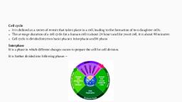
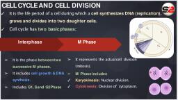
SiC CONCEP, , @ Growth and reproduction are characteristics of cells, in fact of all living organisms, @ Allcells reproduce by dividing into two, with each parental cell giving rise to two daughter cells, each time they divide., 1. Understanding Cell Cycle, , e@ The sequence of events by which a cell duplicates its genome, synthesises the other constituents, of the cell and eventually divides into two daughter cells is termed as cell cycle., , , , @ Although cell growth (in terms of cytoplasmic increase) is a continuous process, DNA synthesis, occurs only during one specific stage in the cell cycle., The replicated chromosomes (DNA) are distributed to daughter nuclei by a complex series of, events during cell division., 2. Phases of Cell Cycle:, e M-phase: The M phase represents the phase when the actual cell division or mitosis occurs., e@ The M phase starts with the nuclear division, corresponding to the separation of daughter, chromosomes (karyokinesis) and usually ends with division of cytoplasm (cytokinesis)., e Interphase: Interphase represents the phase between two successive M phases. The interphase,, though called the resting phase, is the time during which the cell is preparing for division by, undergoing both cell growth and DNA replication in an orderly manner., , , , M (Mitosis), Cell nucieus divides. Pairs of, chromatid separate from each other., , Gp (2nd gap):, Final preparation for mitosis phase. "|, Cell provides energy (ATP) and, synthesizes essential protein, , , , , , »<—«Cytokinesis, Cytoplasm divides to form two new cells., , @9, , G, (1st gap), Cell grows and accumulates, essential protein as well as DNA building block,, , S (Synthesis of DNA), , Cell starts replicating chromosomal DNA., Result in pairs of chromatids, , attached at centrometric region., , , , C}, >, , , , , , Fig. 1.1: Diagrammatic view of the Cell Cycle, , e The interphase is divided into three further phases:, , (i) G, phase (Gap 1): G, phase corresponds to the interval between mitosis and initiation of, DNA replication. During G, phase, the cell is metabolically active and continuously grows, but does not replicate its DNA., , (ii) S phase (Synthesis): S or synthesis phase marks the period during which DNA synthesis or, replication takes place. During this time the amount of DNA per cell doubles. If the initial, amount of DNA is denoted as 2C then it increases to 4C. However, there is no increase, in the chromosome number; if the cell had diploid or 2n number of chromosomes at G,,, then even after S phase the number of chromosomes remains the same, i.e., 2n., , (iii) G, phase (Gap 2): During the Gy phase, proteins are synthesised in preparation for, mitosis while cell growth continues., , , , Cell Cycle and Cell Division | 5
Page 2 :
3. M-phase, , 6, , , , @ Many other cells divide only occasionally as needed to replace cell, that ty, 4 AW Nave je,, of injany on cell death. these cells that do not divide further andl exir ¢ © heen long he, amactive stage called qurercen! age (Gy) of the cell cycle, Gel ,, , Ms in this gta 8 ree Vise |, active but no longer protiferate untess called on to do so depending On they 8 the, &, , tithe, organisa Ment, @ Th animals. mitotic cell division is only seen in the diploid somatic cells wh, , mitotic clivistons are seen in both haploid and diploid cells, tereas jp, , Unis iy known as the mitotic phase, Since the number of « hromosomes, , inthe parent and progeny cells is the same, itis also called as equational /, division, Mitosis is divided into the following four stages:, ® Prophase, , @ Prophase which is the first stage of mitosis follows the §, , and Gy Nd. 4.2: (a) Enrly by,, phases of interphase,, , , , ® Chromosomal material condenses to form compact — mitotic, , chromosomes. Chromosomes are seen to be composed of two, chromatids attached together at the centromere., , , , @ Initiation of the assembly of mitotic spindle, the microtubules,, the proteinaceous components of the cell cytoplasm helps in the FIG. 1.25 (b) Lato Pye, process,, , © Cells at the end of prophase, when viewed under the, , microscope, do not sho, , complexes, endoplasmic reticulum, nucleolus and the nuclear envelope,, , ® Metaphase, © Spindle fibres attach to kinetochores of chromosomes,, @ Chromosomes are moved to spindle, pindle fibres to both poles., , ¢ The plane of alignment of the chromosomes at metaphase is referred to as the r, plate., , equator and get aligned along metaphase plate, , , , Metaphase plate, , Spindle fibers, , Centrioles, , Centromere, Cytosol, Plasma membrane, , , , "Fig. 1.3: Metaphase, , ® Anaphase, , © Centromeres split and chromatids separate., e@ Chromatids move to opposite poles., , , , Fig. 1.4: Anaphase, , Biology—XI: Term-2
Page 3 :
taphase *, , , , @ Telophase:, @ Chromosomes cluster at opposite spindle poles, and their identity ts lost as discrete elements., @ Nuclear envelope assembles around the, chromosome clusters,, @ Nucleolus, Golgi complex and ER reform., , Cytokinesis:, @ Cytokinesis is the physical process of cell division Fig. 1.5: Telophase, which divides the cytoplasm of a parental cell into two daughter cells., , , , @ [nan animal cell, this is achieved by the appearance ofa furrow in the plasma membrane. The, furrow gradually deepens and ultimately joins in the centre dividing the cell cytoplasm into, two., , @ In plant cells, wall formation starts in the centre of the cell and grows outward to meet the, existing lateral walls. The formation of the new cell wall begins with the formation of a simple, , precursor, called the cell-plate that represents the middle lamella between the walls of two, adjacent cells., , Significance of Mitosis:, , @ Mitosis or the equational divi, , , , ion is usually restricted to the diploid cells only., , @ Mitosis results in the production of diploid daughter cells with identical genetic complement, usually., , e@ The growth of multicellular organisms is due to mitosis., , @ Helps in cell repair. The cells of the upper layer of the epidermis, cells of the lining of the gut, and blood cells are being constantly replaced., , @ Mitotic divisions in the meristematic tissues — the apical and the lateral result in a continuous, growth of plants throughout their life., , Meiosi, , @ Meiosis is the phenomenon which occurs in any life cycle that involves the process of sexual, reproduction. The production of offspring by sexual reproduction involves the fusion of two, gametes (each having a complete haploid set of chromosomes)., , @ Thus, meiosis is known to be the specialised form of cell division which reduces the chromosome, number in such a way that each daughter nucleus receives only one set of each kind of, chromosome (ie., maternal and paternal). It results in the production of haploid daughter, , cells. In meiosis, the nucleus divides twice but the replication of chromosome takes place only, once., , , , , , @ The key features of meiosis are as follows:, , (®) Meiosis involves two sequential cycles of nuclear and cell, 11 but only a single cycle of DNA replication., , (2) Meiosis | is initiated after the parental chromosomes have replicated to produce identical, sister chromatids at the S$ phase., , , , ion called meiosis I and meiosis, , () Metosis involves pairing of homologous chromosomes and recombination between them., , () Four haploid cells are formed at the end of meiosis II. Meiotic events can be grouped under, the following phases:, , , , , , , , , , Prophase I Prophase [1, Metaphase I Metaphase I, Anaphase I Anaphase II, Telophase I ‘Telophase I, , , , , , , , , , , Cell Cycle and Cell Division
Page 4 :
7. Meiosis 1, , B > Prophase-I: It is considered to be the most complicated and prolonged ph, s. This phase is further sub-divided into five sub., of chromosomal behaviour:, , ASE as Com, , , , : . : Pared, e Si ar stage mito! ty, the similar stage in Phases on the...”, , basis, , , , e@ Leptotene: known to be the very first stage of meiotic division following the in, , Following features are seen during this phase: iter phase., , (i) Chromosomes becomes gradually visible under light microscope., , (ii) Centrioles start moving towards opposite ends or poles and each centriole dey,, astral rays. relons, , (iii) Each chromosome is attached to the nuclear envelope through the attachment Plate g, both of its ends. eat, , e Zygotene: This is the next sub-stage that takes place after the completion of the Previo, one. This is also a short lived stage like leptotene. Following changes are seen during its, phase:, , (i) Homologous chromosomes pair up. This pairing is done in a such a way that the genes, of the same character present on the two chromosomes lie exactly opposite to each, other. This process of association is known as synapsis., , (ii) Itis revealed from the electron micrographic studies that the formation of synaptonemal, complex takes place by a pair of homologous chromosomes that show synapsis. The, complex so formed on account of synapsis forms a bivalent or a tetrad., , (ii) The number of bivalents are half the total number of chromosomes and are not clearly, visible at this stage. :, , e Pachytene: This is the stage which immediately follows zygotene where the pair of, chromosomes become twisted spirally around each other and cannot be distinguished :, separately. This stage is comparatively long lived as compared to the previous two stages., , Following changes are seen during this stage:, , (i) Bivalent chromosomes clearly seen as tetrads., , (@) In this stage, sometimes exchange of genes or crossing over between the two non-sister, chromatids of homologous chromosomes occurs at the points called recombination 5, nodules, which appear at intervals on synaptonemal complex. By the end of pachytene,, recombination gets completed leaving the chromosomes linked at the sites of crossing over, e Diplotene: It is the stage of longest duration of all. Following changes are observed during, this stage:, , (i) In this the synaptonemal complex appears to get dissolved while the chromatids of each, tetrad remain clearly visible., , (i) Recombined homologous chromosomes of the bivalents get separated and form, chiasmata (X-shaped structures). ., , (iii) Chiasmata formation is necessary for the separation of homologous chromosome which, have undergone the process of crossing-over., , (iv) Diplotene can last for months or years in oocytes of some vertebrates., , @ Diakinesis: This is known to be the final stage of meiotic prophase-I. Also known =, terminalisation due to the shifting of chiasmata towards the end of the chromosome>, Following changes are observed during this stage:, , (i) Chromosomes become fully condensed., (i) Nucleolus degenerates., (ii) Breakdown of nuclear enyelope into vesicles., , 55 igs : fa ates . ‘ we ous, (iv) Formation of meiotic spindle (as in mitosis) in order to prepare the homolog!, , chromosomes for separation,, 8 | Biology—XI: Term-2, , . —
Page 5 :
Metaphase-I: The bivalent chromosomes align on the equatorial plane. The microtubules from, the opposite poles of the spindle attach to the pair of homologous chromosomes., , Anaphase-I: The homologous chromosomes separa, at their centromeres,, , , , ¢ while sister chromatids remain associated, , , , ‘Telophase-I: The nuclear membrane :, called as diad of cells. The, , , , nd nucleolus reappears; cytokinesis follows and this is, ge between the two meiotic divisions is called interkinesis and is, , generally short lived. Interkinesis is followed by prophase II, a much simpler prophase than, prophase 1,, , , , , , i hase I, nara ge, Fig. 1.6: Stages of Melosis |, leiosis IT, Prophase-II: Meiosis II is initiated immediately afier cytokinesis, usually before the, chromosomes have fully elongated. In contrast to meiosis I, meiosis II resembles a normal, mitosis, The nuclear membrane disappears by the end of prophase II. The chromosomes again, become compact., Metaphase II: At this stage, the chromosomes align at the equator and the microtubules from, opposite poles of the spindle get attached to the kinetochores of sister chromatids., Anaphase II: It begins with the simultaneous splitting of the centromere of each chromosome, (which was holding the sister chromatids together), allowing them to move toward opposite, poles of the cell., ‘Telophase-II: This is known to be the last stage of second meiotic division and show changes, equally opposite to that of the prophase-II. Meiosis ends up with the progress of this particular, phase, i-e., telophase-II. Following changes are seen during this process:, e Formation of a nuclear envelope (from ER) around each set of chromosomes., , © Nucleoli reappears due to the synthesis of ribosomal RNA (rRNA) and ribosomal DNA, (rDNA)., , , , Fig. 1.7: Stages of Melosis I!, , Cell Cycle and Cell Division | 9


