Page 1 :
4, , Tissues, , I. Epithelial Tissue 65, Special Characteristics of Epithelia 66, Classification of Epithelia 66, Glands 72, Epithelial Surface Features 74, , II. Connective Tissue 77, Special Characteristics of Connective Tissues 78, Structural Elements of Connective Tissues 78, Classification of Connective Tissues 81, Covering and Lining Membranes 89, , III. Muscle Tissue 93, IV. Nervous Tissue 93, Tissue Response to Injury 95, Inflammation 95, Repair 97, , The Tissues Throughout Life 97, , T, , he cells of the human body do not operate independently of, one another. Instead, related cells live and work together in cell, communities called tissues. A tissue is defined as a group of, cells of similar structure that perform a common function. However,, tissues do not consist entirely of cells: Between the cells is a nonliving, material, the extracellular matrix., The word tissue derives from the Old French word meaning “to, weave,” reflecting the fact that the different tissues are woven together, to form the “fabric” of the human body. The four basic types of tissue, are epithelial tissue, connective tissue, muscle tissue, and nervous, tissue. If a single, broad functional term were assigned to each basic, tissue, the terms would be covering (epithelial tissue), support (connective), movement (muscle), and control (nervous). However, these, terms reflect only a fraction of the functions that each tissue performs., , ▲, , Simple columnar epithelium from the small intestine (colored TEM).
Page 2 :
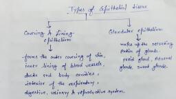
Chapter 4 Tissues 65, Cilia, , Narrow, extracellular, space, Microvilli, , Apical region of, an epithelial cell, , Cell junctions, Tight junction, Epithelium, , Adhesive belt, Desmosome, Gap junction, , Basal region, Basal, lamina, Reticular, fibers, , Basement, membrane, , Nerve ending, Connective, tissue, Capillary, , Figure 4.1 Special characteristics of epithelium. A sheet of closely joined epithelial, cells rests on connective tissue proper. Epithelia contain nerve endings but no blood, vessels. Note the special features on the epithelial cell surfaces: cilia, microvilli, cell, junctions, and basal lamina., , Tissues are the building blocks of the body’s organs., Because most organs contain all four tissue types, learning, about the structure and functions of tissues will provide a, strong foundation for your understanding of the structure and, functions of the organs discussed in the remaining chapters of, this book., , I. EPITHELIAL TISSUE, learning outcomes, ▶ List the functional and structural characteristics of epithelial, tissue., ▶ Identify the different epithelia of the body, and describe, the chief function(s) and location(s) of each., , An epithelium (ep″ ı̆-the′le-um; “covering”) is a sheet, of cells that covers a body surface or lines a body cavity, (Figure 4.1)., , Epithelial tissue occurs in two different forms:, t Covering and lining epithelium covers the outer and inner, , surfaces of most body organs. Examples include the outer, layer of the skin; the inner lining of all hollow viscera,, such as the stomach and respiratory tubes; the lining of the, peritoneal cavity; and the lining of all blood vessels., t Glandular epithelium forms most of the body glands., Epithelia occur at the boundary between two different environments. The epidermis of the skin, for example, lies between, the inside and the outside of the body. Most substances that, enter into the body or are released from the body must pass, through an epithelium. Therefore all functions of epithelia, reflect their roles as interface tissues. These functions include:, 1. Protection of the underlying tissues, 2. Secretion (release of molecules from cells), 3. Absorption (bringing small molecules into cells)
Page 3 :
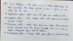
66 Chapter 4 Tissues, , 4. Diffusion (movement of molecules down their concentration gradient), 5. Filtration (passage of small molecules through a sievelike membrane), 6. Sensory reception, , Apical surface, , Basal surface, , Simple, Apical surface, , These functions will be discussed further as we describe the, specific types of epithelia., , Special Characteristics of Epithelia, Epithelial tissues have many characteristics that distinguish, them from other tissue types (Figure 4.1):, 1. Cellularity. Epithelia are composed almost entirely of cells., These cells are separated by a minimal amount of extracellular material, mainly projections of their integral membrane proteins into the narrow spaces between the cells., 2. Specialized contacts. Adjacent epithelial cells are directly, joined at many points by special cell junctions., 3. Polarity. All epithelia have a free apical surface and an, attached basal surface. The structure and function of the, apical and basal surfaces differ, a characteristic called, polarity. The apical surface abuts the open space of a, cavity, tubule, gland, or hollow organ. The basal surface, lies on a thin supporting sheet, the basal lamina, which, is part of the basement membrane (see Figure 4.1; these, structures are further explained on p. 76)., 4. Support by connective tissue. All epithelial sheets in, the body are supported by an underlying layer of connective tissue., 5. Avascular but innervated. Whereas most tissues in the, body are vascular (contain blood vessels), epithelium is, avascular (a-vas′ku-lar), meaning it lacks blood vessels., Epithelial cells receive their nutrients from capillaries in, the underlying connective tissue. Although blood vessels, do not penetrate epithelial sheets, nerve endings do; that, is, epithelium is innervated., 6. Regeneration. Epithelial tissue has a high regenerative, capacity. Some epithelia are exposed to friction, and, their surface cells rub off. Others are destroyed by hostile substances in the external environment such as bacteria, acids, and smoke. As long as epithelial cells receive, adequate nutrition, they can replace lost cells quickly by, mitosis, cell division., , Classification of Epithelia, Many kinds of epithelia exist in the body. Two features are, used to classify and name epithelia: the number of cell layers, and the shape of the cells (Figure 4.2). The terms simple and, stratified describe the number of cell layers in an epithelium, (Figure 4.2a)., t� Simple epithelia contain a single layer of cells, with each, , cell attached to the basement membrane., t� Stratified epithelia contain more than one layer of, cells. The cells on the basal surface are attached to the, , Basal surface, Stratified, (a) Classification based on number of cell layers, , Squamous, , Cuboidal, , Columnar, (b) Classification based on cell shape, , Figure 4.2 Classification of epithelia., , basement membrane; those on the apical surface border, an open space., Cell shape is described as squamous, cuboidal, or columnar,, referring to the appearance of the cells in section (Figure 4.2b)., In each case, the shape of the nucleus conforms to the shape of, the cell. This is an important feature to observe when distinguishing epithelial types., t� Squamous cells (sqwa′mus; “scale”) are flat cells with flat,, , disc-shaped nuclei., t� Cuboidal cells are cube-shaped cells with spherical, centrally located nuclei.
Page 4 :
Chapter 4 Tissues 67, , Table 4.1, , Function of Epithelial Tissue Related to Tissue Type, Number of Layers, , Cell Shape, , One Layer: Simple Epithelial Tissues, , More Than One Layer: Stratified Epithelial Tissues, , Squamous, , Diffusion and filtration, , Protection, , Cuboidal, Columnar, , Secretion and absorption; ciliated types, propel mucus or reproductive cells, , Protection; these tissue types are rare in humans, (see pp. 69–70), , Transitional, , t� Columnar cells are taller than they are wide, like col-, , umns. The nuclei of columnar cells are located near the, basal surface and are commonly oval in shape, elongated, from top to bottom., Simple epithelia are easy to classify by cell shape because, all cells in the layer usually have the same shape. In stratified, epithelia, however, the cell shapes usually differ among the, different cell layers. To avoid ambiguity, stratified epithelia are, named according to the shape of the cells in the apical layer., It is useful to keep in mind how tissue structure reflects tissue function (Table 4.1). Stratified epithelial tissues function to, protect. Multiple layers of cells protect underlying connective, tissues in areas where abrasion is common. In simple epithelia,, the shape of the cells is indicative of tissue function. Squamous, cells are found where diffusion or filtration are important,, because these are distance-dependent processes; the thinner, the layer, the more quickly the process occurs. Columnar and, cuboidal cells are found in tissues involved in secretion and, absorption. Larger cells are necessary for the additional cellular machinery needed to produce and package secretions and to, produce the necessary energy for these processes. Ciliated epithelia function to propel material, for example, mucus. Keep, these generalizations in mind as you study each type of epithelial tissue in detail., As you view micrographs of different types of epithelium, (Figure 4.3), try to pick out the individual cells within each., This is not always easy, because the boundaries between epithelial cells often are indistinct. Furthermore, the nucleus of, a particular cell may or may not be visible, depending on the, plane of the cut made to prepare the tissue slides. When a pear, is sliced in transverse sections, slices from the top of the pear, will not contain any seeds, but slices from the middle region, will. The same is true for sections through tissues: Some cells, may be sliced through the nucleus, whereas others may not., , Simple Epithelia, Simple Squamous Epithelium (Figure 4.3a) A simple, squamous epithelium is a single layer of flat cells. When, viewed from above, the closely fitting cells resemble a tiled, floor. When viewed in lateral section, they resemble fried, , Protection; stretching to accommodate distension, of urinary structures, , eggs seen from the side. Thin and often permeable, this type, of epithelium occurs wherever small molecules pass through, a membrane quickly, by processes of diffusion or filtration., The walls of capillaries consist exclusively of this epithelium,, whose exceptional thinness encourages efficient exchange of, nutrients and wastes between the bloodstream and surrounding tissue cells. In the lungs, this epithelium forms the thin, walls of the air sacs, where gas exchange occurs., , Simple Cuboidal Epithelium (Figure 4.3b) Simple, cuboidal epithelium consists of a single layer of cube-shaped, cells. This epithelium forms the secretory cells of many, glands, the walls of the smallest ducts of glands, and the walls, of many tubules in the kidney. Its functions are the same as, those of simple columnar epithelium., Simple Columnar Epithelium (Figure 4.3c) Simple, columnar epithelium is a single layer of tall cells aligned, like soldiers in a row. It lines the digestive tube from the, stomach to the rectum. It functions in the active movement, of molecules, namely in absorption and secretion. The structure of simple columnar epithelium is ideal for these functions: It is thin enough to allow large numbers of molecules, to pass through it quickly, yet thick enough to house the cellular machinery needed to perform the complex processes of, molecular transport., Some simple columnar epithelia bear cilia (sil′e-ah; “eyelashes”), whiplike bristles on the apex of epithelial cells that, beat rhythmically to move substances across certain body surfaces (see Figure 4.1). A simple ciliated columnar epithelium, lines the inside of the uterine tube. Its cilia help move the ovum, to the uterus. (This journey is traced in Chapter 3.) Cilia are, considered in detail later in this chapter., Pseudostratified Columnar Epithelium (Figure 4.3d), The cells of pseudostratified (soo-do-strat′ı̆-f ı̄d) columnar, epithelium are varied in height. All of its cells rest on the, basement membrane, but only the tall cells reach the apical, surface of the epithelium. The short cells are undifferentiated, and continuously give rise to the tall cells. The cell nuclei, lie at several different levels, giving the false impression that, this epithelium is stratified (pseudo = false)., (Text continues on page 72.)
Page 5 :
68 Chapter 4 Tissues, (a) Simple squamous epithelium, Description: Single layer of flattened cells, with disc-shaped central nuclei and sparse, cytoplasm; the simplest of the epithelia., Air sacs of, lung tissue, Nuclei, of squamous, epithelial, cells, , Function: Allows passage of materials by, diffusion and filtration in sites where protection, is not important; produces lubricating, fluid in serosae., , Location: Kidney glomeruli; air sacs of lungs;, lining of heart, blood vessels, and lymphatic, vessels; lining of ventral body cavity (serosae)., , Photomicrograph: Simple squamous epithelium, forming part of the alveolar (air sac) walls (140×)., , (b) Simple cuboidal epithelium, Description: Single layer of cubelike cells, with large, spherical central nuclei., , Simple, cuboidal, epithelial, cells, , Function: Secretion and absorption., , Basement, membrane, Location: Kidney tubules; ducts and secretory, portions of small glands; ovary surface., Connective, tissue, , Photomicrograph: Simple cuboidal epithelium, in kidney tubules (430×)., , Figure 4.3 Epithelial tissues., , View, , Histology
Page 6 :
Chapter 4 Tissues 69, , (c) Simple columnar epithelium, Description: Single layer of tall cells with, round to oval nuclei; some cells bear cilia;, layer may contain mucus-secreting, unicellular glands (goblet cells)., Microvilli, Goblet cell, Simple, columnar, epithelial, cell, , Function: Absorption; secretion of mucus,, enzymes, and other substances; ciliated type, propels mucus (or reproductive cells) by, ciliary action., Location: Nonciliated type lines most of the, digestive tract (stomach to rectum),, gallbladder, and excretory ducts of some, glands; ciliated variety lines small bronchi,, uterine tubes, and some regions of the uterus., , Basement, membrane, , Photomicrograph: Simple columnar epithelium of the small, intestine (650×)., , (d) Pseudostratified columnar epithelium, Description: Single layer of cells of differing, heights, some not reaching the free surface;, nuclei seen at different levels; may contain, mucus-secreting goblet cells and bear cilia., , Cilia, Goblet cell, , Pseudostratified, epithelial, layer, , Function: Secretion, particularly of mucus;, propulsion of mucus by ciliary action., Location: Nonciliated type in male’s, sperm-carrying ducts and ducts of large, glands; ciliated variety lines the trachea, most, of the upper respiratory tract., , Basement, membrane, Trachea, Photomicrograph: Pseudostratified ciliated columnar, epithelium lining the human trachea (780×)., , Figure 4.3 Epithelial tissues, continued
Page 7 :
70 Chapter 4 Tissues, (e) Stratified squamous epithelium, Description: Thick membrane composed of, several cell layers; basal cells are cuboidal or, columnar and metabolically active; surface, cells are flattened (squamous); in the, keratinized type, the surface cells are full of, keratin and dead; basal cells are active in, mitosis and produce the cells of the more, superficial layers., Stratified, squamous, epithelium, , Function: Protects underlying tissues in, areas subjected to abrasion., Location: Nonkeratinized type forms the, moist linings of the esophagus, mouth, and, vagina; keratinized variety forms the, epidermis of the skin, a dry membrane., , Nuclei, Basement, membrane, Connective, tissue, , Photomicrograph: Stratified squamous epithelium, lining the esophagus (280×)., , (f) Stratified cuboidal epithelium, Description: Generally two, layers of cubelike cells., Basement, membrane, , Cuboidal, epithelial, cells, , Function: Protection., , Location: Largest ducts of sweat glands,, mammary glands, and salivary glands., Duct, lumen, , Photomicrograph: Stratified cuboidal epithelium forming, a salivary gland duct (290×)., , Figure 4.3 Epithelial tissues, continued
Page 8 :
Chapter 4 Tissues 71, , (g) Stratified columnar epithelium, , Description: Several cell layers;, basal cells usually cuboidal;, superficial cells elongated, and columnar., , Stratified, columnar, epithelium, Basement, membrane, Function: Protection; secretion., Location: Rare in the body; small, amounts in male urethra and in large ducts, of some glands., , Underlying, connective, tissue, , Urethra, Photomicrograph: Stratified columnar epithelium lining the, male urethra (360×)., , (h) Transitional epithelium, Description: Resembles both stratified, squamous and stratified cuboidal; basal cells, cuboidal or columnar; surface cells dome, shaped or squamous-like, depending on, degree of organ stretch., , Transitional, epithelium, Function: Stretches readily and permits, distension of urinary organ by contained urine., Location: Lines the ureters, bladder, and part, of the urethra., Basement, membrane, Connective, tissue, Photomicrograph: Transitional epithelium lining the bladder,, relaxed state (365×); note the bulbous, or rounded, appearance, of the cells at the surface; these cells flatten and become, elongated when the bladder is filled with urine., , Figure 4.3 Epithelial tissues, continued
Page 9 :
72 Chapter 4 Tissues, , Pseudostratified columnar epithelium, like simple columnar epithelium, functions in secretion or absorption. A ciliated, type lines the interior of the respiratory tubes. Here, the cilia, propel sheets of dust-trapping mucus out of the lungs., , Stratified Epithelia, Stratified epithelia contain two or more layers of cells. They, regenerate from below; that is, the basal cells divide and push, apically to replace the older surface cells. Stratified epithelia, are more durable than simple epithelia, and their major (but, not only) role is protection., , Stratified Squamous Epithelium (Figure 4.3e) As, you might expect, stratified squamous epithelium consists, of many cell layers whose surface cells are squamous. In, the deeper layers, the cells are cuboidal or columnar. Of all, the epithelial types, this is the thickest and best adapted for, protection. It covers the often-abraded surfaces of our body,, forming the epidermis of the skin and the inner lining of the, mouth, esophagus, and vagina. To learn the location of stratified squamous epithelium, simply remember that this epithelium forms the outermost layer of the skin and extends a, certain distance into every body opening that is directly continuous with the skin., The epidermis of the skin is keratinized, meaning that, its surface cells contain an especially tough protective protein called keratin. The other stratified squamous epithelia, of the body lack keratin and are nonkeratinized. (Keratin is, explained in detail in Chapter 5, p. 105)., Stratified Cuboidal and Columnar Epithelia (Figure 4.3f, Stratified cuboidal and stratified columnar, epithelia are rare types of tissue, located in the large ducts of, some glands, for example, sweat glands, mammary glands,, and salivary glands. Stratified columnar epithelium is also, found in small amounts in the male urethra., , and Figure 4.3g), , Transitional Epithelium (Figure 4.3h) Transitional epithelium lines the inside of the hollow urinary organs. Such, organs (the urinary bladder, for example) stretch as they, fill with urine. As the transitional epithelium stretches, it, thins from about six cell layers to three, and its apical cells, unfold and flatten. When relaxed, portions of the apical surface invaginate into the cell, giving this surface a scalloped, appearance. Thus, this epithelium undergoes “transitions” in, shape. It also forms an impermeable barrier that keeps urine, from passing through the wall of the bladder., check your understanding, ◻ 1. Describe the location of the apical region of an, epithelial tissue. How can this region be easily, identified in a micrograph?, ◻ 2. Why are cuboidal or columnar cells found in epithelia, that function in secretion and absorption? List three, locations of cuboidal epithelium., ◻ 3. Why is columnar epithelium not found in locations, where diffusion occurs? What type of epithelium is, found in these locations?, (For answers, see Appendix B.), , Glands, learning outcomes, ▶ Distinguish exocrine from endocrine glands., ▶ Explain how multicellular exocrine glands are classified., , Epithelial cells that make and secrete a product form glands., The products of glands are aqueous (water-based) fluids that, usually contain proteins. Secretion is the process whereby, gland cells obtain needed substances from the blood and, transform them chemically into a product that is then discharged from the cell. More specifically, the protein product, is made in the rough endoplasmic reticulum (ER), then packaged into secretory granules by the Golgi apparatus and ultimately released from the cell by exocytosis (see p. 28 and, Figure 2.8). These organelles are well developed in most, gland cells that secrete proteins., Glands are classified as endocrine (en′do-krin; “internal, secretion”) or exocrine (ek′so-krin; “external secretion”),, depending on where they release their product, and as, unicellular (“one-celled”) or multicellular (“many-celled”), on the basis of cell number. Unicellular glands are scattered within epithelial sheets, whereas most multicellular, glands develop by invagination of an epithelial sheet into, the underlying connective tissue., , Endocrine Glands, Endocrine glands lack ducts, so they are often referred to as, ductless glands. They secrete directly into the tissue fluid that, surrounds them. More specifically, endocrine glands produce, messenger molecules called hormones (hor′mōnz; “exciters”), which they release into the extracellular space. These, hormones then enter nearby capillaries and travel through the, bloodstream to specific target organs, which are commonly far, removed from the endocrine gland that produces the hormone., Each hormone signals its target organs to respond in some, characteristic way. For example, endocrine cells in the intestine secrete a hormone that signals the pancreas to release the, enzymes that help digest a meal., Although most endocrine glands derive from epithelia,, some derive from other tissues. (The endocrine system is discussed in detail in Chapter 17.), , Exocrine Glands, Exocrine glands are numerous, and many of their products, are familiar ones. All exocrine glands secrete their products onto body surfaces (skin) or into body cavities (such, as the digestive tube), and multicellular exocrine glands, have ducts that carry their product to the epithelial surfaces. The activity of an exocrine secretion is local; that, is, the secretion acts near the area where it is released., Exocrine glands are a diverse group: They include many, types of mucus-secreting glands, the sweat glands and oil, glands of the skin, salivary glands of the mouth, the liver, (which secretes bile), the pancreas (which secretes digestive enzymes), mammary glands (which secrete milk), and, many others.
Page 10 :
Chapter 4 Tissues 73, , Microvilli, , Secretory, vesicles, containing, mucin, , Golgi, apparatus, Rough ER, , Nucleus, , (a), , (b), , Figure 4.4 Goblet cell (unicellular exocrine gland). (a) Photomicrograph of a, mucus-secreting goblet cell in the simple columnar epithelium that lines the small, intestine (1650* ). (b) Diagram of a goblet cell. Note the secretory vesicles and the, well-developed rough ER and Golgi apparatus., , Unicellular Exocrine Glands The only important example, of a one-celled exocrine gland is the goblet cell (Figure 4.4)., True to its name, a goblet cell is indeed shaped like a goblet,, a drinking glass with a stem. Goblet cells are scattered within, the epithelial lining of the intestines and respiratory tubes,, between columnar cells with other functions. They produce, mucin (mu′sin), a glycoprotein (sugar protein) that dissolves, in water when secreted. The resulting complex of mucin and, water is viscous, slimy mucus. Mucus covers, protects, and, lubricates many internal body surfaces., Multicellular Exocrine Glands Each multicellular exocrine gland has two basic parts: an epithelium-walled duct, and a secretory unit consisting of the secretory epithelium, (Figure 4.5). Also, in all but the simplest glands, a supportive connective tissue surrounds the secretory unit, carrying, with it blood vessels and nerve fibers. Often, the connective, tissue forms a fibrous capsule that extends into the gland, proper and partitions the gland into subdivisions called, lobes (not illustrated)., , Multicellular glands are classified by the structure of, their ducts (Figure 4.5). Simple glands have an unbranched, duct, whereas compound glands have a branched duct., The glands are further categorized by their secretory units:, They are tubular if their secretory cells form tubes and, alveolar (al-ve′o-lar) if the secretory cells form spherical sacs (alveolus = a small, hollow cavity). Furthermore,, some glands are tubuloalveolar; that is, they contain, both tubular secretory units and alveolar units. The term, acinar (as′ı̆-nar; acinus = grape or berry) is commonly used, as a synonym for alveolar., check your understanding, ◻ 4. What type of gland are goblet cells? What do they, secrete?, ◻ 5. An ________ gland secretes into the tissue fluid, and its, secretion is carried to a target organ by the bloodstream., ◻ 6. What feature distinguishes a simple exocrine gland, from a compound exocrine gland?, (For answers, see Appendix B.)
Page 11 :
74 Chapter 4 Tissues, , Simple duct structure, , Compound duct structure, , (duct does not branch), , (duct branches), , Tubular, secretory, structure, Simple tubular, , Simple branched tubular, , Example, Intestinal glands, , Example, Stomach (gastric), glands, , Compound tubular, Example, Duodenal glands of small intestine, , Alveolar, secretory, structure, Simple alveolar, , Simple branched alveolar, , Example, No important example, in humans, , Example, Sebaceous (oil) glands, , Surface epithelium, , Duct, , Compound alveolar, , Compound tubuloalveolar, , Example, Mammary glands, , Example, Salivary glands, , Secretory epithelium, , Figure 4.5 Types of multicellular exocrine glands. Multicellular glands are classified, according to the structure of their ducts (simple or compound) and the structure of, their secretory units (tubular, alveolar, tubuloalveolar)., , Epithelial Surface Features, learning outcome, ▶ Describe apical, lateral, and basal surface features of, epithelia and epithelial cells., , As previously described, epithelial tissues are composed, of many cells closely joined together by special cell junctions along their lateral walls. Epithelial tissues also have, distinct apical and basal regions. The basal surface sits on, a specialized boundary with the underlying connective tissue; the apical region of certain epithelia has modifications, associated with specific functions. These special features, are described next., , Lateral Surface Features: Cell Junctions, Three factors act to bind epithelial cells to one another:, (1) adhesion proteins in the plasma membranes of the, adjacent cells link together in the narrow extracellular space;, (2) the wavy contours of the membranes of adjacent cells join, in a tongue-and-groove fashion; and (3) there are special cell, , junctions (Figure 4.6). Cell junctions, the most important of, the factors, are characteristic of epithelial tissue but are found, in other tissue types as well., , Tight Junctions In the apical region of most epithelial, tissues, a beltlike junction extends around the periphery of, each cell (Figure 4.6a). This is a tight junction, or a zonula, occludens (zōn′u-lah o-klood′enz; “small zone that shuts off ”)., At tight junctions, the adjacent cells are so close that some, proteins in their plasma membranes are fused. This fusion, forms a seal that closes off the extracellular space; thus tight, junctions prevent molecules from passing between the cells, of epithelial tissue. For example, the tight junctions in the, epithelium lining the digestive tract keep digestive enzymes,, ions, and microorganisms in the intestine from seeping into, the bloodstream. Tight junctions need not be entirely impermeable; some are more leaky than others and may let certain, types of ions through., Adhesive Belt Junctions Just below the tight junctions, in epithelial tissues are adhesive belt junctions, or zonula
Page 12 :
Chapter 4 Tissues 75, , Plasma membranes, of adjacent cells, , Microvilli, , Intercellular, space, , Basement membrane, , Intercellular, space, Plaque, , Intercellular, space, , Channel, between cells, (connexon), Interlocking, junctional, proteins, Intercellular, space, , Intermediate, filament, (keratin), (a) Tight junctions: Impermeable junctions, prevent molecules from passing through, the intercellular space., , Linker, glycoproteins, (cadherins), , (b) Desmosomes: Anchoring junctions bind, adjacent cells together and help form an, internal tension-reducing network of fibers., , (c) Gap junctions: Communicating junctions, allow ions and small molecules to pass, from one cell to the next for intercellular, communication., , Figure 4.6 Cell junctions. An epithelial cell shown joined to adjacent cells by three, common types of cell junction., , adherens (zōn′u-lah ad-hir-ens), a type of anchoring junction, (Figure 4.1). Transmembrane linker proteins attach to the, actin microfilaments of the cytoskeleton and bind adjacent, cells. This junction reinforces the tight junctions, particularly, when the tissues are stretched. Together with tight junctions,, these form the tight junctional complex around the apical lateral borders of epithelial tissues., , Desmosomes The main junctions for binding cells, together are called desmosomes (dez′mo-sōmz; “binding, bodies”), an anchoring junction. These adhesive spots are, scattered along the abutting sides of adjacent cells (Figure, 4.6b). Desmosomes have a complex structure: On the cytoplasmic face of each plasma membrane is a circular plaque., The plaques of neighboring cells are joined by linker proteins., These project from both cell membranes and interdigitate,, , like the teeth of a zipper, in the extracellular space. In addition,, intermediate filaments (the cytoskeletal elements that resist, tension) insert into each plaque from its inner, cytoplasmic, side. Bundles of these filaments extend across the cytoplasm, and anchor at other desmosomes on the opposite side of the, same cell. Overall, this arrangement not only holds adjacent, cells together but also interconnects intermediate filaments of, the entire epithelium into one continuous network of strong, guy-wires. The epithelium is thus less likely to tear when, pulled on, because the pulling forces are distributed evenly, throughout the sheet., Desmosomes are found in cardiac muscle tissue as, well as in epithelial tissues. In general, these junctions, are common in tissues that experience great mechanical, stress.
Page 13 :
76 Chapter 4 Tissues, , Gap Junctions A gap junction, or nexus (nek′sus;, “bond”), is a tunnel-like junction that can occur anywhere, along the lateral membranes of adjacent cells (Figure 4.6c)., Gap junctions function in intercellular communication by, allowing small molecules to move directly between neighboring cells. At such junctions, the adjacent plasma membranes are very close, and the cells are connected by hollow, cylinders of protein (connexons). Ions, simple sugars, and, other small molecules pass through these cylinders from, one cell to the next. Gap junctions are common in embryonic tissues and in many adult tissues, including connective, tissues. They are also prevalent in smooth and cardiac muscle, where the passage of ions through gap junctions synchronizes contraction., , Microvillus, , Actin, filaments, , Figure 4.7 Microvilli., , Basal Feature: The Basal Lamina, At the border between the epithelium and the connective tissue deep to it is a supporting sheet called the basal lamina, (lam′ ı̆-nah; “sheet”) (see Figure 4.1). This thin, noncellular sheet consists of proteins secreted by the epithelial cells., Functionally, the basal lamina acts as a selective filter; that, is, it determines which molecules from capillaries in the, underlying connective tissue are allowed to enter the epithelium. The basal lamina also acts as scaffolding along which, regenerating epithelial cells can migrate. Luckily, infections, and toxins that destroy epithelial cells usually leave the basal, lamina in place, for without this lamina, epithelial regeneration is more difficult., Directly deep to the basal lamina is a layer of reticular, fibers (defined shortly) belonging to the underlying connective tissue. Together, these reticular fibers plus the basal, lamina form the basement membrane (see Figure 4.1). The, thin basal lamina can be seen only by electron microscopy,, but the thicker basement membrane is visible by light microscopy (see Figure 4.3). Although this text distinguishes basal, lamina from basement membrane, many scientists use these, two terms interchangeably., CLINICAL APPLICATION, Basement Membranes and Diabetes In untreated, cases of diabetes mellitus (type 1 or type 2 diabetes),, the basement membranes of the epithelial lining of, capillaries thicken over time. This thickening is, caused by increased amounts of glucose, present in, high concentrations in diabetics, binding to the, proteins of the basement membrane. This process, is referred to as increased glycosylation of the basement, membrane. Thickening is especially evident in the, capillaries in the kidneys and retina of the eye, which, can become nonfunctional. For this reason, kidney, failure and blindness are major symptoms of advanced, diabetes., , Apical Surface Features: Microvilli and Cilia, Microvilli (mi″cro-v ı̆′li; “little shaggy hairs”) are fingerlike, extensions of the plasma membrane of apical epithelial cells, , (Figure 4.7). Each microvillus contains a core of actin, filaments that extend into the actin microfilaments of, the cytoskeleton and function to stiffen the microvillus., Microvilli occur on almost every moist epithelium in the, body but are longest and most abundant on epithelia that, absorb nutrients (in the small intestine) (see Figure 4.3c) or, transport ions (in the kidney). In such epithelia, microvilli, maximize the surface area across which small molecules, enter or leave cells. Microvilli are also abundant on epithelia that secrete mucus, where they help anchor the mucous, sheets to the epithelial surface., Cilia are whiplike, highly motile extensions of the apical, surface membranes of certain epithelial cells (see Figure 4.1)., Each cilium contains a core of microtubules held together by, cross-linking and radial proteins (Figure 4.8). The microtubules are arranged in pairs, called doublets, with nine outer, doublets encircling one central pair. Ciliary movement is, generated when adjacent doublets grip one another with side, arms made of the motor protein dynein (p. 33) and these arms, start to oscillate. This causes the doublets to slide along the, length of each other, like centipedes trying to run over each, other’s backs. As a result, the cilum bends., The microtubules in cilia are arranged in much the, same way as in the cytoplasmic organelles called centrioles, (p. 33). Indeed, cilia originate as their microtubules assemble, around centrioles that have migrated from the centrosome, to the apical plasma membrane. The centriole at the base of, each cilium is called a basal body (Figure 4.8a)., The cilia on an epithelium bend and move in coordinated, waves, like waves across a field of grass on a windy day. These, waves push mucus and other substances over the epithelial surface (Figure 4.8c). Each cilium executes a propulsive power, stroke, followed by a nonpropulsive recovery stroke similar to, feathering an oar or a canoe paddle (Figure 4.8b). This sequence, ensures that fluid is moved in one direction only. An extremely, long, isolated cilium is called a flagellum (flah-jel′-um;, “whip”). The only flagellated cells in the human body are, sperm, which use their flagella to swim through the female, reproductive tract.
Page 14 :
Chapter 4 Tissues 77, Outer microtubule, doublet, Central, microtubule, , Power, or, propulsive, stroke, , The doublets also, have attached, motor proteins,, the dynein arms., , Recovery stroke, when cilium, is returning to its initial position, , Dynein arms, , Cross-linking, proteins inside, outer doublets, , The outer, microtubule, doublets and the, two central, microtubules are, held together by, cross-linking, proteins and, radial spokes., , Radial spoke, Plasma, membrane, , 1, , 2, , 3, , 4, , 5, , 6, , 7, , (b) Phases of ciliary motion, Layer of mucus, , Cilium, , Cell surface, , Basal body, (centriole), (a) Structure of a cilium, (c) Traveling wave created by the activity of, many cilia acting together propels mucus, across cell surfaces., , Figure 4.8 Cilia structure and function., , CLINICAL APPLICATION, Kartagener’s Syndrome A type of immotile cilia, syndrome, Kartagener’s syndrome is an inherited disease, in which the dynein arms within the cilia fail to form., This condition leads to frequent respiratory infections, because the nonfunctional cilia cannot sweep inhaled, bacteria out of the respiratory tubes., , check your understanding, ◻ 7. How do cilia differ from microvilli?, ◻ 8. How do the intermediate filaments within epithelial, cells function to bind together the entire sheet of, epithelia?, ◻ 9. In addition to epithelial tissue, what other types of, tissues contain gap junctions?, (For answers, see Appendix B.), , II. CONNECTIVE TISSUE, learning outcomes, ▶ Describe the features that are common to all connective, tissues., ▶ Identify the four main classes of connective tissue., , The second of the four basic types of tissue is connective, tissue, the most diverse and abundant type of tissue. There, are four main classes of connective tissue and many subclasses. (Table 4.2, p. 79). The main classes are:, 1. Connective tissue proper, familiar examples of which are, fat tissue and the fibrous tissue of ligaments, 2. Cartilage, 3. Bone tissue, 4. Blood, Connective tissues do far more than just connect the tissues, and organs of the body together. They also form the basis, of the skeleton (bone and cartilage), store and carry nutrients (fat tissue and blood), surround all the blood vessels and, nerves of the body (connective tissue proper), and lead the, body’s fight against infection., In this section, we will first discuss the special characteristics of connective tissues and then describe the structural, elements found in connective tissues. Finally, we will review, the structure, function, and location of the specific types of, connective tissues.
Page 15 :
78 Chapter 4 Tissues, Extracellular, matrix, , Cell types, , Ground substance, Macrophage, , Fibers, Collagen fiber, Elastic fiber, Reticular fiber, , Fibroblast, , Lymphocyte, , Fat cell, , Capillary, , Mast cell, , Neutrophil, , Figure 4.9 Areolar connective tissue: A model connective tissue., , Special Characteristics, of Connective Tissues, As different as they are, fat, bone, and blood are all connective, tissues. All connective tissues share the same simple structural, plan (Figure 4.9)., 1. Relatively few cells, lots of extracellular matrix. The, cells of connective tissues are separated from one, another by a large amount of extracellular material, called the extracellular matrix (ma′triks; “womb”)., This differs markedly from epithelial tissue, whose, cells crowd closely together., 2. Extracellular matrix composed of ground substance, and fibers. The extracellular matrix is produced by the, cells of the connective tissue. It is composed of some, type of ground substance embedded with protein fibers., The ground substance varies for each class of connective tissue. In many, it is a soft, gel-like substance that, holds tissue fluid. In bone, it is hard—calcified by inorganic calcium salts. The fibrous portion of the matrix, provides support for the connective tissue. Three types, of protein fibers are found in connective tissues: collagen fibers, reticular fibers, and elastic fibers. The types,, density, and distribution of the fibers are distinctive for, each type of connective tissue. The differences among, , Practice art labeling, , the physical properties and functions of each type of, connective tissue are due to differences in the composition of the extracellular matrix. The details of the matrix, structure will be discussed with each type of connective, tissue., 3. Embryonic origin. Another feature common to connective tissues is that they all originate from the embryonic, tissue called mesenchyme (Figure 4.10; see also p. 51)., , Structural Elements, of Connective Tissues, Connective tissues are composed of cells and a significant, amount of extracellular matrix. Connective tissues differ in, their physical properties because of differences in the types of, cells and the composition of the extracellular matrix. However,, they all share similar structural elements: cells, fibers, and, ground substance., , Cells, In most connective tissues, the primary cell type produces, the extracellular matrix (see Table 4.2, third column). In, connective tissue proper, these cells are called fibroblasts, (literally, “fiber buds” or “fiber formers”). Fibroblasts make, the protein subunits of fibers, which are secreted into the, extracellular matrix and assemble into fibers. Fibroblasts
Page 16 :
Chapter 4 Tissues 79, , Table 4.2, , Comparison of Classes of Connective Tissues, Components, , Tissue Class, and Example, , Subclasses, , Cells, , Matrix, , General Features, , Connective Tissue, Proper, , 1. Loose connective, tissue, , Fibroblasts, , Gel-like ground substance, , Fibrocytes, , All three fiber types:, collagen, reticular, elastic, , Six different types; vary in, density and types of fibers, , t� "SFPMBS, , Defense cells, , t� "EJQPTF, , Fat cells, , Functions as a binding, tissue, Resists mechanical stress,, particularly tension, , t� 3FUJDVMBS, 2. Dense connective, tissue, t� 3FHVMBS, Dense regular, connective tissue, , Cartilage, , t� MSSFHVMBS, t� &MBTUJD, 1. Hyaline cartilage, 2. Elastic cartilage, 3. Fibrocartilage, , Chondroblasts, found in growing, cartilage, Chondrocytes, , Gel-like ground, substance, Fibers: collagen, elastic, fibers in some, , Resists compression, because of the large, amounts of water held in, the matrix, Functions to cushion and, support body structures, , Hyaline cartilage, Bone Tissue, , 1. Compact bone, , Osteoblasts, , 2. Spongy bone, , Osteocytes, , Gel-like ground, substance calcified with, inorganic salts, , Hard tissue that resists both, compression and tension, Functions in support, , Fibers: collagen, , Compact bone, Blood, , Blood cell formation, and differentiation, are quite complex, (Details are provided, in Chapter 18), , Erythrocytes (RBCs), , Plasma, , A fluid tissue, , Leukocytes (WBCs), , No fibers, , Functions to carry O2, CO2,, nutrients, wastes, and other, substances (hormones, for, example), , Platelets, , also secrete the molecules that form the ground substance, of the matrix. In cartilage tissue, the cells that secrete the, matrix are chondroblasts (kon′dro-blasts; “cartilage formers”), and in bone they are osteoblasts (os″te-o-blasts′;, “bone formers”). Once these tissue-forming cells are not, actively secreting new matrix, they are termed fibrocytes,, chondrocytes, and osteocytes (cyte = cell). They function, , to maintain and repair the tissue matrix and keep the tissue, healthy., The cells found in blood are an exception. These cells, do not produce the plasma matrix of blood. The cellular, components of blood function to carry respiratory gases, (red blood cells), to fight infections (white blood cells), and, to aid in blood clotting (platelets).
Page 17 :
80 Chapter 4 Tissues, , Additional cell types are found in many connective tissues (shown in Figure 4.9)., t� Fat cells, or adipose (ad′ı̆-p ōs) cells, store nutrients. Fat, , cells are egg-shaped, and their cytoplasm is dominated, by a single, giant lipid droplet that flattens the nucleus, and cytoplasm at one end of the cell. Mature fat cells are, among the largest cells in the body and cannot divide., t� White blood cells (neutrophils, lymphocytes, eosinophils), respond to and protect against infectious agents., t� Macrophages specialize in phagocytosis. They engulf and, devour a wide variety of foreign materials, ranging from, whole bacteria to foreign molecules, dirt particles, and, dead tissue cells., t� Mast cells mediate inflammation and promote healing., Mast cells contain many large secretory granules filled, with numerous chemicals that are secreted in response, to infectious and allergy-inducing agents. The chemicals, secreted by mast cells include histamine (increases blood, flow and capillary permeability in the local area), heparin, (interacts with other mast cell chemicals), and proteases, (protein-degrading enzymes)., , Fibers, The extracellular matrix of a connective tissue is composed, of fibers and ground substance (Figure 4.9). Three types of, fibers are found in connective tissues: collagen fibers, reticular fibers, and elastic fibers. In general, the fibers function in, support, yet each type of fiber contributes unique properties, to the connective tissue., Collagen fibers (shown as thick, whitish gray fibers in, Figures 4.9 and 4.10), are the strongest and most abundant, type of fiber in connective tissues. Collagen fibers resist tension (pulling forces) and contribute strength to a connective, tissue. Pulling tests show that collagen fibers are stronger, than steel fibers of the same size! The thick collagen fibers, that one sees with the light microscope are bundles of thinner, collagen fibrils, which consist of still thinner strands that are, strongly cross-linked to one another. This cross-linking is the, source of collagen’s great tensile strength., Reticular (re-tik′u-lar) fibers (shown as thin blue, fibers in Figures 4.9 and 4.10), are bundles of a special type, of collagen fibril. These short fibers cluster into a meshlike, network (reticulum = network) that covers and supports, the structures bordering the connective tissue. For example,, capillaries are coated with fuzzy nets of reticular fibers,, and these fibers form part of the basement membrane of, epithelia. Individual reticular fibers glide freely across one, another when the network is pulled, so they allow more give, than collagen fibers do., The last fiber type found in certain connective tissues is, elastic fibers (illustrated as gold fibers in Figures 4.9 and, 4.10). Elastic fibers contain the rubberlike protein elastin, (e-last′in), which allows them to function like rubber bands., Although we typically think of rubber bands as being, stretchy, the defining feature of an elastic material is its ability to recoil back to its original form after being stretched., , Connective tissue can stretch only so much before its thick,, ropelike collagen fibers become taut. When the tension is, released, the elastic fibers recoil, and the stretched tissue, resumes its original shape., CLINICAL APPLICATION, Scurvy Vitamin C, which is abundant in, citrus fruits such as oranges and lemons, is necessary, for the proper cross-linking of the molecules that, make up collagen fibers. A deficiency of this vitamin, in the diet can lead to scurvy, a weakening of collagen, and connective tissue throughout the body. Strong, collagen is necessary for holding teeth in their, sockets, reinforcing the walls of blood vessels, healing, wounds, and forming scar tissue. Common signs of, scurvy include loss of teeth, blood vessel rupture, and, poor healing. Scurvy was a common ailment of sea, explorers in the sixteenth to eighteenth centuries. In, the mid-1700s, a British naval physician observed that, consumption of citrus juice prevented scurvy. For that, reason, the British Royal Navy began serving lime juice to, sailors to prevent the disease—and British sailors came to, be called “limeys.”, , Ground Substance, The other component of the matrix of connective tissue is the, ground substance (Figure 4.9). The molecules that compose, the ground substance are produced and secreted by the primary, cell type of the connective tissue (fibroblasts, chondroblasts, or, osteoblasts). The ground substance in most connective tissues, is a gel-like material that consists of large sugar and sugarprotein molecules (proteoglycans and glycosaminoglycans)., These molecules soak up fluid like a sponge. The fluid-filled, ground substance functions to cushion and protect body structures (connective tissue proper), to withstand compressive, stresses (cartilage), or to hold the tissue fluid that bathes all the, cells in our body (areolar connective tissue). In bone tissue, the, secreted ground substance is embedded with calcified mineral, salts, which make the matrix hard and contribute to its function, in supporting the body., Here again, blood is an exception. The ground substance, of blood, plasma, is not produced by the blood cells. Blood, plasma is composed of water (about 90%) and various dissolved proteins, nutrients, ions, gases, and other molecules., These molecules are either produced by cells in other organs, and then secreted into blood (for example, plasma proteins, and hormones) or transported into blood from an external, source (water, air, and nutrients)., A model of connective tissue can be built with a few simple ingredients: gelatin, jellybeans, and licorice. Dissolve the, gelatin, and allow it to set. The gelatin represents the ground, substance of the connective tissue. Gelatin is composed, of proteins (actually hydrolyzed collagen) that hold fluid,, very similar to the ground substance of most connective tissues. Once the gelatin thickens (approximately 1.5 hours or, so), add a few jellybeans and some licorice. The jellybeans, represent the cellular components of connective tissue, and
Page 18 :
Chapter 4 Tissues 81, , licorice represents the fibers. Different types of licorice can, represent different types of fibers: thick red twists represent, collagen fibers; thin black laces represent elastic fibers. This, model will not mimic the physical properties of connective, tissues—strength, elasticity, and resistance to compression—, but it does give you a visual analogy of the composition of a, connective tissue., check your understanding, ◻ 10. How do epithelial tissues differ from connective, tissues in the arrangement of the cells in each tissue?, ◻ 11. Distinguish the matrix of a connective tissue from the, ground substance., ◻ 12. Which structural element of connective tissue resists, tension? Which resists compression? Which allows for, recoil? Which element produces the matrix?, (For answers, see Appendix B.), , Classification of Connective Tissues, learning outcome, ▶ Differentiate the different types of connective tissues in, reference to the types of cells located within the tissue,, the structure and composition of the extracellular matrix,, and the main function of each., , Connective Tissue Proper—Loose, Connective Tissues, Connective tissue proper has two subclasses: loose connective, tissue (areolar, adipose, and reticular) and dense connective, tissue (dense irregular, dense regular, and elastic). These subclasses are distinguished by the density of fibers: In loose connective tissues, the fibers are distributed throughout the tissue, but are separated from each other by ground substance. There, are three types of loose connective tissue: areolar, adipose,, and reticular., , Areolar Connective Tissue (Figure 4.9 and Figure 4.10b), Loose areolar connective tissue is the most widespread type, of connective tissue proper. This tissue underlies almost all, the epithelia of the body and surrounds almost all the small, nerves and blood vessels, including the capillaries. The structure of this tissue reflects its basic functions:, 1., 2., 3., 4., , Supporting and binding other tissues, Holding body fluids, Defending the body against infection, Storing nutrients as fat, , The fibers of areolar connective tissue provide support., Areolar connective tissue has all three types of fibers in its, extracelluar matrix: collagen fibers, reticular fibers, and elastic, fibers (although the reticular fibers do not show up when the, common histological stains hematoxylin and eosin are used)., The ground substance of areolar connective tissue holds, fluid. All the cells of the body are bathed in tissue fluid, or, interstitial fluid. This fluid is derived from leakage of fluid, and small molecules from the blood as it travels through the, , capillaries. Nutrients and oxygen are delivered to cells and, waste molecules are carried away from cells via diffusion, through this fluid. Areolar connective tissue lies between the, capillaries and all other cells and tissues in the body. It soaks, up this tissue fluid, much like a sponge. Thus it keeps the, body’s cells surrounded by fluid and facilitates the passage of, nutrients, gases, waste products, and other molecules to and, from the cells., Areolar connective tissue is the body’s first line of, defense against invading microorganisms, such as bacteria,, viruses, fungi, and parasites. Lying just deep to the epithelial tissues that line the body surfaces and surrounding capillaries, it is in an ideal location to destroy microorganisms, at their entry site—before they enter the capillaries and use, the vascular system to spread to other locations. Areolar connective tissue contains a variety of defense cells (shown in, Figure 4.9), which respond to infectious agents., Cellular defenses are not the only means by which areolar connective tissue fights infection. The viscous ground substance and the dense networks of collagen fibers in the extracellular matrix slow the progress of invading microorganisms., Some bacteria, however, secrete enzymes that rapidly break, down ground substance or collagen. Such matrix-degrading, bacteria are highly invasive; that is, they spread rapidly through, the connective tissues and are especially difficult for the body’s, defenses to control. An example of such a bacterium is the, Streptococcus strain responsible for strep throat., A minor function of areolar connective tissue is to store, energy reserves as fat. Fat cells occur singly or in small, groups in this tissue (see Figure 4.9)., , Adipose Tissue (Figure 4.10c) Adipose tissue is similar, to areolar connective tissue in structure and function, but, its nutrient-storing function is much greater. Correspondingly,, adipose tissue is crowded with fat cells, which account for, 90% of its mass. These fat cells are grouped into large clusters, called lobules. Adipose tissue is richly vascularized, reflecting, its high metabolic activity. It removes lipids from the bloodstream after meals and later releases them into the blood, as, needed. Without the fat stores in our adipose tissue, we could, not live for more than a few days without eating., Much of the body’s adipose tissue occurs in the layer, beneath the skin called the hypodermis. Adipose tissue also, is abundant in the mesenteries, which are sheets of serous, membranes that hold the stomach and intestines in place. Fat, in this location is called visceral fat. Additionally, fat forms, cushioning pads around the kidneys and behind the eyeballs, in the orbits., Whereas the abundant fat beneath the skin serves the, general nutrient needs of the entire body, smaller depots of, fat serve the local nutrient needs of highly active organs., Such depots occur around the hard-working heart and around, lymph nodes (where cells of the immune system are furiously, fighting infection), within some muscles, and as individual, fat cells in the bone marrow (where new blood cells are produced at a frantic rate). Many of these local depots offer special lipids that are highly enriched., (Text continues on page 84.)
Page 19 :
82 Chapter 4 Tissues, (a) Embryonic connective tissue: mesenchyme, , Description: Embryonic connective tissue;, gel-like ground substance containing fibers;, star-shaped mesenchymal cells., , Mesenchymal, cells, , Ground, substance, , Function: Gives rise to all other connective, tissue types., , Fibers, , Location: Primarily in embryo., , Photomicrograph: Mesenchyme, an embryonic connective, tissue (385×). The matrix is composed of the fluid ground, substance (clear-appearing background) and fine, sparse fibers., , (b) Connective tissue proper: loose connective tissue, areolar, Description: Gel-like matrix with all three fiber, types; cells: fibroblasts, macrophages, mast, cells, and some white blood cells., Elastic, fibers, , Ground, substance, Function: Wraps and cushions organs; its, macrophages phagocytize bacteria; plays, important role in inflammation; holds and, conveys tissue fluid., , Fibroblast, nuclei, , Location: Widely distributed under epithelia, of body, e.g., forms lamina propria of mucous, membranes; packages organs; surrounds, capillaries., , Collagen, fibers, , Epithelium, , Lamina, propria, , Figure 4.10 Connective tissues., , Photomicrograph: Areolar connective tissue, a soft packaging, tissue of the body (340×)., , View, , Histology
Page 20 :
Chapter 4 Tissues 83, , (c) Connective tissue proper: loose connective tissue, adipose, Description: Matrix as in areolar connective, tissue, but very sparse; closely packed, adipocytes, or fat cells, have nucleus pushed, to the side by large fat droplet., , Nucleus of, fat cell, , Function: Provides reserve food fuel;, insulates against heat loss; supports and, protects organs., Vacuole, containing, fat droplet, , Location: Under skin in the hypodermis;, around kidneys and eyeballs; within abdomen;, in breasts., Adipose, tissue, , Mammary, glands, , Photomicrograph: Adipose tissue from the subcutaneous, layer under the skin (350×)., , (d) Connective tissue proper: loose connective tissue, reticular, Description: Network of reticular fibers in a, typical loose ground substance; reticular cells, lie on the network., , White blood cell, (lymphocyte), , Function: Fibers form a soft internal skeleton, (stroma) that supports other cell types, including white blood cells, mast cells, and, macrophages., Location: Lymphoid organs (lymph nodes,, bone marrow, and spleen)., , Spleen, , Figure 4.10 Connective tissues, continued., , Reticular, fibers, , Photomicrograph: Dark-staining network of reticular connective, tissue fibers forming the internal skeleton of the spleen (350×).
Page 21 :
84 Chapter 4 Tissues, (e) Connective tissue proper: dense connective tissue, dense irregular, Description: Primarily irregularly arranged, collagen fibers; some elastic fibers; major cell, type is the fibroblast; defense cells and fat, cells are also present., , Nuclei of, fibroblasts, , Function: Able to withstand tension exerted, in many directions; provides structural, strength., Location: Fibrous capsules of organs and, of joints; dermis of the skin; submucosa of, digestive tract., , Fibrous, layer of, joint capsule, , Collagen, fibers, , Photomicrograph: Dense irregular connective tissue from the, dermis of the skin (300×)., , Figure 4.10 Connective tissues, continued., , The typical, nutrient-storing fat is white adipose tissue, or white fat. Another type, called brown adipose, tissue, produces heat and is a nutrient consumer. Brown fat,, thought to occur only in babies to aid in thermoregulation,, has also been identified in adults. It is located in the hypodermis between the two scapulae (shoulder blades) in the, center of the back, on the side of the anterior neck, and on, the anterior abdominal wall. It is even more richly vascularized than white fat. Each brown-fat cell contains many lipid, droplets and numerous mitochondria, which use the lipid, fuel to heat the bloodstream rather than to produce ATP, molecules., , Reticular Connective Tissue (Figure 4.10d) Reticular, connective tissue resembles areolar tissue, but the only, fibers in its matrix are reticular fibers. These fine fibers, form a broad, three-dimensional network like the frame of, a house. The spaces in the framework create a labyrinth of, caverns that hold many free cells. The bone marrow, spleen,, and lymph nodes, which house many free blood cells outside, their capillaries, consist largely of reticular connective tissue., Fibroblasts called reticular cells lie along the reticular network of this tissue. (Reticular tissue is discussed further in, Chapters 18 and 21.), , Connective Tissue Proper—Dense, Connective Tissue, Dense connective tissue, or fibrous connective tissue, contains, more collagen than areolar connective tissue does. With its, thick collagen fibers, it can resist extremely strong pulling (tensile) forces. There are three types of dense connective tissue:, irregular, regular, and elastic., , Dense Irregular Connective Tissue (Figure 4.10e), Dense irregular connective tissue is similar to areolar tissue, but its collagen fibers are much thicker. These, fibers run in different planes, allowing this tissue to resist, strong tensions from different directions. This tissue dominates the leathery dermis of the skin, which is commonly, stretched, pulled, and hit from various angles. This tissue, also makes up the fibrous capsules that surround certain, organs in the body, such as kidneys, lymph nodes, and, bones. Its cellular and matrix elements are the same as in, areolar connective tissue., Dense irregular connective tissue is confusing, even for, experts. The dermis obviously fits the name because its fibers, run randomly, as one would expect for a tissue called “irregular.” However, the fibrous capsules of organs consist of two, (Text continues on page 86.)
Page 22 :
Chapter 4 Tissues 85, , (f) Connective tissue proper: dense connective tissue, dense regular, Description: Primarily parallel collagen fibers;, a few elastic fibers; major cell type is the, fibroblast., , Collagen, fibers, , Function: Attaches muscles to bones or to, muscles; attaches bones to bones; withstands, great tensile stress when pulling force is, applied in one direction., , Nuclei of, fibroblasts, , Location: Tendons, most, ligaments, aponeuroses., , Shoulder, joint, Ligament, , Photomicrograph: Dense regular connective tissue from a, tendon (425×)., , Tendon, , (g) Connective tissue proper: dense connective tissue, elastic, Description: Dense regular connective tissue, containing a high proportion of elastic fibers., , Function: Allows recoil of tissue following, stretching; maintains pulsatile flow of blood, through arteries; aids passive recoil of lungs, following inspiration., , Elastic fibers, , Location: Walls of large arteries; within, certain ligaments associated with the vertebral, column; within the walls of the bronchial tubes., , Aorta, Heart, , Figure 4.10 Connective tissues, continued., , Photomicrograph: Elastic connective tissue in the wall of the, aorta (250×).
Page 23 :
86 Chapter 4 Tissues, (h) Cartilage: hyaline, Description: Amorphous but firm matrix;, collagen fibers form an imperceptible network;, chondroblasts produce the matrix and, when, mature (chondrocytes), lie in lacunae., Chondrocyte, in lacuna, , Function: Supports and reinforces; serves as, resilient cushion; resists compressive stress., , Matrix, , Location: Forms most of the embryonic, skeleton; covers the ends of long bones in, joint cavities; forms costal cartilages of the, ribs; cartilages of the nose, trachea, and, larynx., , Costal, cartilages, , Photomicrograph: Hyaline cartilage from a costal cartilage, of a rib (470×)., , Figure 4.10 Connective tissues, continued., , layers, with all the fibers in a layer running parallel to each, other but perpendicular to the fibers in the other layer. That, is, two perpendicular “regular” layers can make the tissue, “irregular.”, , Dense Regular Connective Tissue (Figure 4.10f) All, collagen fibers in dense regular connective tissue usually run in the same direction, parallel to the direction of, pull. Crowded between the collagen fibers are rows of fibroblasts, which continuously manufacture the fibers and a scant, ground substance. When this tissue is not under tension, its, collagen fibers are slightly wavy. Unlike areolar connective, tissue, dense regular connective tissue is poorly vascularized, and contains no fat cells or defense cells., With its enormous tensile strength, dense regular connective tissue is the main component of ligaments, bands, or sheets that bind bones to one another. It also is the main, tissue in tendons, which are cords that attach muscles to, bones, and aponeuroses (ap″o-nu-ro′s ēs), which are sheetlike tendons., Dense regular connective tissue also forms fascia, (fash′e-ah; “a band”), a fibrous membrane that wraps around, muscles, muscle groups, large vessels, and nerves. Many, sheets of fascia occur throughout the body, binding structures together like plastic sandwich wrap. When the word, fascia is used alone, it is understood to mean deep fascia., , Superficial fascia, something else entirely, is the fatty hypodermis below the skin., , Elastic Connective Tissue (Figure 4.10g) In elastic, connective tissue, elastic fibers are the predominant type of, fiber, and bundles of elastic fibers outnumber the bundles of, collagen fibers. This tissue is located in structures where recoil, from stretching is important: within the walls of arteries, in certain ligaments (ligamentum nuchae and ligamentum flavum,, which connect successive vertebrae), and surrounding the bronchial tubes in the lungs., check your understanding, ◻ 13. How does loose connective tissue differ from dense, connective tissue?, ◻ 14. Which type of connective tissue forms the following, structures: ligaments and tendons; the hypodermis, of the skin; the tissue that underlies epithelia; lymph, nodes?, (For answers, see Appendix B.), , Cartilage, As you have seen, connective tissue proper has the ability to resist tension (pulling). Cartilage and bone are the, (Text continues on page 89.)
Page 24 :
Chapter 4 Tissues 87, , (i) Cartilage: elastic, Description: Similar to hyaline cartilage, but, more elastic fibers in matrix., , Chondrocyte, in lacuna, , Function: Maintains the shape of a structure, while allowing great flexibility., Matrix, , Location: Supports the external ear, (pinna); epiglottis., , Photomicrograph: Elastic cartilage from the human ear pinna;, forms the flexible skeleton of the ear (510×)., , (j) Cartilage: fibrocartilage, Description: Matrix similar to but less firm, than that in hyaline cartilage; thick collagen, fibers predominate., , Function: Tensile strength with the ability to, absorb compressive shock., Collagen, fibers, , Location: Intervertebral discs; pubic, symphysis; discs of knee joint., Intervertebral, discs, , Chondrocytes, in lacunae, , Photomicrograph: Fibrocartilage from an intervertebral disc, (175×)., , Figure 4.10 Connective tissues, continued.
Page 25 :
88 Chapter 4 Tissues, (k) Others: bone (osseous tissue), Description: Hard, calcified matrix containing, many collagen fibers; osteocytes lie in, lacunae. Very well vascularized., , Central, canal, Lacunae, , Function: Supports and protects, (by enclosing); provides levers for the muscles, to act on; stores calcium and other minerals, and fat; marrow inside bones is the site for, blood cell formation (hematopoiesis)., , Lamella, , Location: Bones., , Photomicrograph: Cross-sectional view of bone (175×)., , (l) Connective tissue: blood, Description: Red and white blood cells in a, fluid matrix (plasma)., Red blood, cells, (erythrocytes), , White blood cells:, r�-ZNQIPDZUF, r�/FVUSPQIJM, Function: Transport respiratory gases,, nutrients, wastes, and other substances., , Location: Contained within blood vessels., Plasma, , Photomicrograph: Smear of human blood (1650×); shows two, white blood cells surrounded by red blood cells., , Figure 4.10 Connective tissues, continued.
Page 26 :
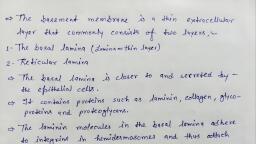
Chapter 4 Tissues 89, , firm connective tissues that resist compression (pressing) as, well as tension. Like all connective tissues, they consist of, cells separated by a matrix containing fibers, ground substance, and tissue fluid. However, these skeletal tissues exaggerate the supportive functions of connective tissue and play, no role in fat storage or defense against disease., Cartilage (Figure 4.10h–j), a firm but flexible tissue, occurs in several parts of the skeleton. For example,, it forms the supporting rings of the trachea (windpipe) and, gives shape to the nose and ears. Like nearly all connective, tissues, cartilage consists of cells separated by an abundant, extracellular matrix (Figure 4.10h). This matrix contains, thin collagen fibrils, a ground substance, and an exceptional, quantity of tissue fluid; in fact, cartilage consists of up to, 80% water! The arrangement of water in its matrix enables, cartilage to spring back from compression (as explained in, Chapter 6, p. 126.), Cartilage is simpler than other connective tissues: It, contains no blood vessels or nerves and just one kind of, cell, the chondrocyte. Each chondrocyte resides within, a cavity in the matrix called a lacuna. Immature chondrocytes are chondroblasts, cells that actively secrete the, matrix during cartilage growth. Cartilage is found in three, varieties, each dominated by a particular fiber type: hyaline cartilage, elastic cartilage, and fibrocartilage. (These, are introduced in Figure 4.10 h–j and are also discussed in, Chapter 6, pp. 125–127.), , Bone, Because of its rocklike hardness, bone tissue has a tremendous ability to support and protect body structures. Bone, matrix contains inorganic calcium salts, which enable, bone to resist compression, and an abundance of collagen, fibers, which allow bone to withstand strong tension, (Figure 4.10k, p. 88)., Immature bone cells, called osteoblasts, secrete the collagen fibers and ground substance of the matrix. Then calcium, salts precipitate on and between the collagen fibers, hardening, the matrix. The mature bone cells, called osteocytes, inhabit, cavities (lacunae; singular: lacuna) in this hardened matrix., Bone is a living and dynamic tissue, well supplied with blood, vessels. (It is discussed further in Chapter 6.), , Blood, Blood (Figure 4.10l), the fluid in the blood vessels, is the, most atypical connective tissue. It does not bind things, together or give mechanical support. It is classified as a connective tissue because it develops from mesenchyme and, consists of blood cells surrounded by a nonliving matrix, the, liquid blood plasma. Its cells and matrix are very different, from those in other connective tissues. Blood functions as, the transport vehicle for the cardiovascular system, carrying, defense cells, nutrients, wastes, respiratory gases, and many, other substances throughout the body. (Blood is discussed in, detail in Chapter 18.), Epithelial and connective tissues are the most, diverse tissue types in the body. For this reason, they are, , commonly the most difficult for students to understand. Focus on Distinguishing Between Epithelial and, Connective Tissues (Figure 4.11) presents the distinctions, between these types of tissues in reference to structure, and function., check your understanding, ◻ 15. Which connective tissues contain collagen fibers?, ◻ 16. In which connective tissues are the cells located, in lacunae?, ◻ 17. Which component of cartilage tissue functions, to resist compressive forces?, (For answers, see Appendix B.), , Covering and Lining Membranes, learning outcome, ▶ Discuss the structure and function of mucous, serous, and, cutaneous membranes., , Now that you have learned about connective and epithelial tissues, you can consider the covering and lining membranes, that combine these two tissue types (Figure 4.12). These, membranes, which cover broad areas within the body, consist, of an epithelial sheet plus the underlying layer of connective, tissue proper. These membranes are of three types: cutaneous,, mucous, and serous., The cutaneous membrane (ku-ta′ne-us; “skin”) is the, skin, covering the outer surface of the body (Figure 4.12a). Its, outer epithelium is the thick epidermis, and its inner connective tissue is the dense dermis. It is a dry membrane. (The skin, is discussed further in Chapter 5.), A mucous membrane, or mucosa (mu-ko′sah), lines, the inside of every hollow internal organ that opens to the, outside of the body. More specifically, mucous membranes, line the tubes of the respiratory, digestive, reproductive, and, urinary systems (Figure 4.12b). Although different mucous, membranes vary widely in the types of epithelia they contain, all are wet or moist. As their name implies, many, mucous membranes secrete mucus. Not all of them do so,, however., All mucous membranes consist of an epithelial sheet, directly underlain by a layer of loose areolar connective tissue called the lamina propria (lam′ ı̆-nah pro′pre-ah; “one’s, own layer”). In the mucous membranes of the digestive system, the lamina propria rests on a layer of smooth muscle, cells. Later chapters discuss the specific mucous membranes, of the body., Serous membranes, or serosae (introduced in Chapter 1),, are the slippery membranes that line the closed pleural, pericardial, and peritoneal cavities (Figures 4.12c and 1.7, p. 12)., A serous membrane consists of a simple squamous epithelium, called mesothelium, lying on a thin layer of areolar, connective tissue. This membrane produces a slippery serous, fluid, beginning as a filtrate of the blood in capillaries in the, connective tissue, with the addition of lubricating molecules, by the mesothelium.
Page 27 :
FOCUS, , Identifying Epithelial and Connective Tissues, , Figure 4.11 The structure of a tissue provides clues to its name and location., How are the cells distributed in epithelial tissue?, Lots of cells, little extracellular material, , EPITHELIAL TISSUES, , Tightly packed cells, , Epithelia form selective barriers for the passage of, molecules across body surfaces. Epithelia line the, external and internal surfaces of many organs and, form the secretory portion of most glands., , Apical and basal regions, Apical region borders open space, Basal region attached to, underlying connective tissue, , How are the cell nuclei arranged?, , Single row of nuclei:, , Stacked rows of nuclei:, , Cell heights differ, nuclei, appear stacked; each cell, attached to basal region:, , Simple epithelium, , Stratified epithelium, , Pseudostratified epithelium, , What shape are the cells at the apical region?, Squamous Simple squamous epithelium, , Stratified squamous epithelium, , Thin, flat cells, , Squamous, cells in apical, region, , Elongated, nucleus, , Multiple layers, of cells, , Thin layer of cells allows for rapid transport of, molecules. Found where diffusion and, filtration occur (see Figure 4.3a)., , Multiple layers of cells provide protection, and rapid regeneration. Found in areas that, experience abrasion or friction (see Figure, 4.3e)., , Cuboidal, , Simple cuboidal epithelium, , Square- to, circularshaped cells, , Stratified cuboidal epithelium, Cuboidal cell, in apical region, , Circular nucleus, in center of cell, Larger cells house cellular organelles active, in secretion and absorption yet allow rapid, passage through epithelial layer (see Figure, 4.3b)., , Columnar, , Simple columnar epithelium, , Rectangularshaped cells, , Two layers of cells protect underlying tissue, from glandular secretions. Forms the ducts of, many large glands (see Figure 4.3f)., , Columnar, cell in apical, region, , Oval nucleus, Elongated columnar cells house cellular, organelles necessary for a tissue active in, secretion and absorption (see Figure 4.3c)., , 90, , Pseudostratified, columnar epithelium, , Stratified columnar epithelium, , Two layers of cells protect underlying, tissues; columnar cells have space for, organelles needed to produce secretions, (see Figure 4.3g)., , Cilia, Goblet, cell, Columnar, cell, Columnar cells and goblet cells produce, secretions; cilia move fluid along the, epithelial surface (see Figure 4.3d).
Page 28 :
View, , Histology, , How are the cells distributed in connective tissue?, Few cells, widely spaced apart, , CONNECTIVE TISSUES (CT), , Fibroblast nuclei, , Connective tissues are binding and support, tissues. They provide structural support, connect, body structures, store nutrients, and hold tissue, fluids. The physical properties of each connective, tissue result from the composition of its matrix., , Lots of extracellular matrix, Fibers, Ground substance, , Where are the cells located within the matrix?, , Cells in direct contact, with matrix components:, , Cells in spaces (lacunae), within the extracellular matrix:, , Cells suspended, in a fluid matrix:, , Connective tissue proper, , Bone and Cartilage, , Blood, , Bone, , Cartilage, , Calcified ground, substance, , Gelatinous ground, substance, Leukocyte, , Loose CT, , Dense CT, , Loosely dispersed, individual fibers, , Densely packed, bundles of fibers, , Osteocytes, in lacunae, Extracellular, matrix, , Areolar CT, Fibroblast, nucleus, , Dense irregular CT, Fibroblast, nuclei, , Extracellular matrix contains, collagen fibers and calcified ground, substance. Strong, stiff, composite, tissue functions in support and, protection (see Figure 4.10k)., , Thick, collagen, fibers, , Fibers, , Erythrocytes, Platelets, , Fluid matrix transports nutrients,, signaling molecules, and waste, products. Cellular components, transport respiratory gases and, fight infections (see Figure, 4.10l)., , Hyaline cartilage, Chondrocyte, , Dense regular CT, , Adipose CT, Fat cell, nuclei, , Fibroblast, nuclei, , Lacuna, Extracellular, matrix, , Elastic cartilage, , Extracellular, matrix, , Thick, collagen, fibers, , Chondrocyte, Lacuna, , Elastic CT, , Reticular CT, , Elastic, fibers, , Fibrocartilage, Fibers, , Matrix holds fluid, binds and, supports. Cells fight, infections and store nutrients., Loose CTs fill body spaces, and allow for exchange of, materials with the blood (see, Figure 4.10b–d)., , Thick, elastic, fibers, Thick collagen fibers resist tension;, elastic fibers enable recoil., Alignment of fibers indicates the, direction of applied forces. Dense, CTs are strong, resilient binding, tissues (see Figure 4.10 e–g)., , Chondrocyte, in lacuna, Collagen, fibers, Firm, resilient matrix. Ground, substance holds fluid and resists, compression; fibers add tensile, strength (collagen) and elasticity, (elastic). Cartilage contributes to, flexible support (see Figure 4.10h–j)., , 91
Page 29 :
92 Chapter 4 Tissues, (c) Serous membranes, , (a) Cutaneous membrane, The cutaneous membrane (the skin) covers the body surface., , Serous membranes line body cavities, that are closed to the exterior., , Cutaneous, membrane, (skin), , Parietal, pleura, Visceral, pleura, , Parietal, pericardium, , (b) Mucous membranes, , Visceral, pericardium, , Mucous membranes line body cavities, that are open to the exterior., , Mucosa of, nasal cavity, Mucosa of, mouth, Esophagus, lining, , Mucosa of, lung bronchi, , Parietal, peritoneum, Visceral, peritoneum, , Figure 4.12 Types of membranes.
Page 30 :
Chapter 4 Tissues 93, , (a) Skeletal muscle, Description: Long, cylindrical, multinucleate, cells; obvious striations., Striations, , Function: Voluntary movement; locomotion;, manipulation of the environment; facial, expression., , Nuclei, , Location: In skeletal muscles attached to, bones or occasionally to skin., Part of, muscle, fiber (cell), , Photomicrograph: Skeletal muscle (450×)., Notice the obvious banding pattern and the, fact that these large cells are multinucleate., , Figure 4.13 Muscle tissues., , III. MUSCLE TISSUE, learning outcome, ▶ Briefly describe the three types of muscle tissue., , The two remaining tissue types are muscle and nervous tissues. These are sometimes called composite tissues because,, along with their own muscle or nerve cells, they contain small, amounts of areolar connective tissue. (Areolar connective tissue, surrounds all blood vessels, and both muscle and nervous tissue, are richly vascularized.), Muscle tissues (Figure 4.13) bring about most kinds, of body movements. Most muscle cells are called muscle, fibers. They have an elongated shape and contract forcefully as they shorten. These cells contain many myofilaments (mi″o-fil′ah-ments; “muscle filaments”), cellular, organelles filled with the actin and myosin filaments that, bring about contraction in all cell types (p. 32). There are, three kinds of muscle tissue: skeletal, cardiac, and smooth., Skeletal muscle tissue (Figure 4.13a) is the major component of organs called skeletal muscles, which pull on, bones to cause body movements. Skeletal muscle cells are, long, large cylinders that contain many nuclei. Their obvious, striated, or banded, appearance reflects a highly organized, arrangement of their myofilaments. (Skeletal muscle tissue is, described in detail in Chapter 10.), , View, , Histology, , Cardiac muscle tissue (Figure 4.13b) occurs in the wall, of the heart. It contracts to propel blood through the blood, vessels. Like skeletal muscle cells, cardiac muscle cells are, striated. However, they differ in two ways: (1) Each cardiac, cell has just one nucleus, and (2) cardiac cells branch and join, at special cellular junctions called intercalated (in-ter′kah-la″ted) discs. (The details of cardiac muscle tissue are discussed, in Chapter 19.), Smooth muscle tissue (Figure 4.13c) is so named, because there are no visible striations in its cells. These cells, are elongated with tapered ends and contain one centrally, located nucleus. Smooth muscle primarily occurs in the walls, of hollow viscera such as the digestive and urinary organs,, uterus, and blood vessels. It generally acts to squeeze substances through these organs by alternately contracting and, relaxing. (Smooth muscle tissue is described in detail in, Chapter 23.), , IV. NERVOUS TISSUE, learning outcome, ▶ Distinguish the cell types found in nervous tissue., , Nervous tissue is the main component of the nervous, organs—the brain, spinal cord, and nerves—which regulate, and control body functions. It contains two types of cells,
Page 31 :
94 Chapter 4 Tissues, (b) Cardiac muscle, Description: Branching, striated, generally, uninucleate cells that interdigitate at, specialized junctions (intercalated discs)., , Striations, Function: As it contracts, it propels blood, into the circulation; involuntary control., , Intercalated, discs, , Location: The walls of the heart., , Nucleus, , Photomicrograph: Cardiac muscle (355×);, notice the striations, branching of cells, and, the intercalated discs., , (c) Smooth muscle, Description: Spindle-shaped cells with, central nuclei; no striations; cells arranged, closely to form sheets., , Smooth, muscle, cell, Function: Propels substances or objects, (foodstuffs, urine, a baby) along internal, passageways; involuntary control., , Nuclei, Location: Mostly in the walls of hollow, organs., , Photomicrograph: Sheet of smooth muscle from the, digestive tract (465×)., , Figure 4.13 Muscle tissues, continued.
Page 32 :
Chapter 4 Tissues 95, , Nervous tissue, Description: Neurons are branching, cells; cell processes that may be quite, long extend from the nucleus-containing, cell body; also contributing to nervous, tissue are nonconducting supporting cells,, neuroglia (not illustrated)., Neuron processes, , Cell body, of a neuron, , Cell body, Neuron, processes, , Axon Dendrites, , Function: Transmit electrical signals, from sensory receptors and to effectors, (muscles and glands) that control, the activity of the effector organs., , Nuclei of, neuroglia, , Location: Brain, spinal, cord, and nerves., , Photomicrograph: Neurons (125×)., , Figure 4.14 Nervous tissue., , neurons and supporting cells. Neurons are the highly specialized nerve cells (Figure 4.14) that generate and conduct, electrical impulses. They have extensions, or processes, that, allow them to transmit impulses over substantial distances, within the body. Dendrites are cell processes that extend, from the cell body of a neuron like branches of a tree. The, dendrites, the receptive region of the neuron, transmit signals, toward the cell body. A neuron also has a singular, long cell, process extending from its cell body, the axon, that generates, nerve impulses and transmits them away from the cell body., The supporting cells, called neuroglia, are nonconducting, cells that nourish, insulate, and protect the delicate neurons., (A more complete discussion of nervous tissue appears in, Chapter 12.), check your understanding, ◻ 18. Look at a photomicrograph of smooth muscle tissue, (Figure 4.13c). How can you tell this tissue is not, stratified squamous epithelium?, ◻ 19. Which nerve cells function to transmit electrical, impulses?, ◻ 20. Distinguish between the terms striated and stratified., Which tissues are striated? Which tissues are, stratified?, (For answers, see Appendix B.), , View, , Histology, , TISSUE RESPONSE TO INJURY, learning outcome, ▶ Describe the inflammatory and repair processes by which, tissues recover from injury., , The body has many mechanisms for protecting itself from, injury and invading microorganisms. Intact epithelia act, as a physical barrier, but once that barrier has been penetrated, protective responses are activated in the underlying, connective tissue proper. These are the inflammatory and, immune responses. Inflammation is a nonspecific, local, response that develops quickly and limits the damage to the, injury site. The immune response, by contrast, takes longer to, develop and is highly specific. It destroys particular infectious, microorganisms and foreign molecules at the site of infection, and throughout the body. This section concentrates on inflammation. (The immune response is discussed in Chapter 21.), , Inflammation, Almost every injury or infection leads to an inflammatory, response. For example, assume your skin is cut by a dirty piece, of glass, crushed by a blow in football, or has an infected pimple. As short-term, or acute, inflammation develops in the, connective tissue, it produces four symptoms: heat, redness,, swelling, and pain. You can trace the source of each symptom.
Page 34 :
Chapter 4 Tissues 97, , At first glance, inflammatory edema seems detrimental, but it is actually beneficial. The entry of blood-derived, fluid into the injured connective tissue (1) helps to dilute, toxins secreted by bacteria; (2) brings in oxygen and, nutrients from the blood, necessary for tissue repair, and, (3) brings in antibodies from the blood to fight infection., In very severe infections and in all wounds that sever blood, vessels, the fluid leaking from the capillaries contains, clotting proteins. In these cases, clotting occurs in the connective tissue matrix. The fibrous clot isolates the injured, area and “walls in” the infectious microorganisms, preventing their spread., The next stage in inflammation is stasis (“standing”)., This is the slowdown in local blood flow that necessarily, follows a massive exit of fluid from the capillaries. At this, stage, white blood cells begin to leave the small vessels. First, to appear at the infection site are neutrophils, then macrophages. These cells devour the infectious microorganisms, and the damaged tissue cells as well., , cellular division occurs in both these tissues after damage, and, efforts are under way to coax them to regenerate better., In nonregenerating tissues and in exceptionally severe, wounds, fibrosis totally replaces the lost tissue. The resulting, scar appears as a pale, often shiny area and shrinks during the, months after it first forms. A scar consists mostly of collagen, fibers and contains few cells or capillaries. Although it is very, strong, it lacks the flexibility and elasticity of most normal, tissues, and it is unable to perform the normal functions of the, tissue it has replaced., Irritation of visceral organs can cause them to adhere to one, another or to the body wall as they scar. Such adhesions can, prevent the normal churning actions of loops of the intestine,, dangerously halting the movement of food through the digestive, tube. They can also restrict the movement of the heart and lungs, and immobilize the joints. After almost all abdominal surgeries,, adhesions form between the body wall and the abdominal viscera, making subsequent surgery in that region more difficult., , Repair, , ◻ 21. Which tissues regenerate easily? Which tissues do, not regenerate?, ◻ 22. Is the scar tissue that creates a “scar” located in the, epithelium or in the underlying connective tissue?, ◻ 23. What causes the heat and swelling in an infected tissue?, , Even as inflammation proceeds, repair begins. Tissue repair, can occur in two major ways: by regeneration and by fibrosis. Regeneration is the replacement of a destroyed tissue by, new tissue of the same kind, whereas fibrosis involves the, proliferation of a fibrous connective tissue called scar tissue., Tissue repair in a skin wound involves both regeneration and, fibrosis. After the blood within the cut has clotted, the surface, part of the clot dries to form a scab (Figure 4.15, 1 ). At this, point, the repair begins with a step called organization., Organization is the process by which the clot is replaced, by granulation tissue (Figure 4.15, 2 ). Granulation tissue, is a delicate pink tissue made of several elements. It contains, capillaries that grow in from nearby areas, as well as proliferating fibroblasts that produce new collagen fibers to bridge, the gash. Some of its fibroblasts have contractile properties, that pull the margins of the wound together. As organization proceeds, macrophages digest the original clot, and the, deposit of collagen continues. As more collagen is made, the, granulation tissue gradually transforms into fibrous scar tissue (Figure 4.15, 3 )., During organization, the surface epithelium begins to, regenerate, growing under the scab until the scab falls away., The end result is a fully regenerated epithelium and an underlying area of scar., The process we have just outlined describes the healing of, a wound such as a cut, scrape, or puncture. In pure infections, (a pimple or sore throat), by contrast, there is usually no clot, formation or scarring. Only severe infections lead to scarring., The capacity for regeneration varies widely among the, different tissues. Epithelia regenerate extremely well, as do, bone, areolar connective tissue, dense irregular connective, tissue, and blood-forming tissue. Smooth muscle and dense, regular connective tissue have a moderate capacity for regeneration, but skeletal muscle and cartilage have only a weak, capacity. Cardiac muscle and the nervous tissue in the brain, and spinal cord have no functional regenerative capacity., However, recent studies have shown that some unexpected, , check your understanding, , (For answers, see Appendix B.), , THE TISSUES THROUGHOUT LIFE, learning outcome, ▶ Indicate the embryonic derivation of each tissue class., ▶ Briefly describe changes that occur in tissues with age., , The embryonic derivations of the four basic tissues are as, follows (Figure 4.16):, t� Connective and muscle tissue derive from mesenchyme,, , mostly of the mesoderm germ layer., t� Most epithelial tissues develop from embryonic epithelium of the ectoderm and endoderm layers. A few, epithelia—namely, the epithelium lining the vessels and, the mesothelium lining the ventral body cavity—derive, from mesodermal mesenchyme., t� Nervous tissue in the brain and spinal cord derives from, ectodermal epithelium (recall Chapter 3, p. 52)., By the end of the second month of development, the, four primary tissues have formed, and all major organs are, in place. In virtually all tissues, the cells continue to divide, throughout the prenatal period, providing the rapid body, growth that occurs before birth. The division of nerve cells,, however, stops or nearly stops during the fetal period. After, birth, the cells of most other tissues continue to divide until, adult body size is reached. In adulthood, only the epithelial, tissues and blood cell–forming tissues are highly mitotic., Cellular division in other tissues slows greatly, although, many tissues retain a regenerative capacity.
Page 35 :
98 Chapter 4 Tissues, , 16-day-old embryo, (dorsal surface view), Muscle and connective tissue, (mostly from mesoderm), , Ectoderm, , Epithelium, (from all three, germ layers), , Mesoderm, Endoderm, , Nervous tissue, (from ectoderm), , Inner lining of, digestive and respiratory, organs (from endoderm), , Figure 4.16 Embryonic germ layers and the primary tissue types they produce., , Some tissues that regenerate throughout life do so, through the division of their mature, differentiated cells., This is common in epithelial tissues. The new differentiated, cells replace older cells within the tissue. Abnormalities in, this process can result in pathological conditions. Cancer, is a disease in which tissue cells divide uncontrollably and, expand beyond the normal tissue boundaries. The most, common types of cancers in humans are carcinomas, cancer, arising from epithelia. (See A CLOSER LOOK.), Many tissues contain populations of stem cells, relatively, undifferentiated cells that renew themselves continually and, divide to produce new tissue cells as needed. Stem cells have, long been known to exist in the rapidly replacing tissues,, such as the epidermis, the lining of the digestive tube, some, connective tissues, and blood-forming tissue. Now, however,, they have been found elsewhere: in the brain, adipose tissue,, and probably the liver and pancreas., Given good nutrition, good circulation, and relatively, infrequent wounds and infections, the tissues normally function well through youth and middle age. The importance of, nutrition for tissue health cannot be overemphasized. For, , example, vitamin A is needed for the normal regeneration, of epithelium (liver and carrots are rich in this vitamin)., Because proteins are the structural material of the body, an, adequate intake of protein is essential for the tissues to retain, their structural integrity., With increasing age, the epithelia thin and are more easily breached. The amount of collagen in the body declines,, making tissue repair less efficient. Wounds do not heal as, fast as in youth. Bone, muscle, and nervous tissues begin to, atrophy. These events are due in part to a decrease in circulatory efficiency, which reduces the delivery of nutrients to, the tissues, but in some cases diet is a contributing factor. A, diet low in protein and vitamins can negatively affect tissue, health., check your understanding, ◻ 24. Which embryonic layer or layers form epithelium?, ◻ 25. How do cancerous cells differ from other highly, mitotic cell types?, (For answers, see Appendix B.), , RELATED CLINICAL TERMS, Adenoma (ad-ĕ-no′mah) (aden = gland; oma = tumor) Any, neoplasm of glandular epithelium, benign or malignant. The, malignant type is more specifically called adenocarcinoma., , Carcinoma (kar″sı̆-no′mah) (karkinos = crab, cancer) Cancer, arising in an epithelium. Ninety percent of all human cancers, are of this type: lung, breast, prostate, colon, and others., , Epithelial–Connective Tissue Interactions An exchange of, chemical signals between epithelia and the underlying connective tissues, especially in the control of the events of embryonic, development and in the generation of cancer as epithelial, tumor cells gain the ability to metastasize. These exchanges, , are also called epithelial-stromal interactions or, in embryos,, epithelial-mesenchymal interactions. This is currently a very, active area of research., , Lesion (le′zhun; “wound”) Any injury, wound, or infection that, affects tissue over an area of a definite size, as opposed to being, widely spread throughout the body., , Sarcoma (sar-ko′mah) (sarkos = flesh; oma = tumor) Cancer, arising in the mesenchyme-derived tissues; that is, in connective tissues and muscle.
Page 36 :
A CLOSER LOOK, , Cancer—The Intimate Enemy, Although once perceived as disorganized cell growth, cancer is now, known to be a coordinated process in, which a precise sequence of tiny alterations changes a normal cell into a, killer. Let’s take a closer look at what, cancer really is., When cells fail to honor normal, controls of cell division and multiply, excessively, an abnormal mass of, proliferating cells called a neoplasm, (ne′o-plazm, “new growth”) results., Neoplasms are classified as benign, (“kindly”) or malignant (“bad”). A, benign neoplasm, commonly called a, tumor, is strictly a local affair. Its cells, remain compacted, are often encapsulated, tend to grow slowly, and seldom, kill their hosts if they are removed before they compress vital organs., In contrast, cancers are malignant, neoplasms, nonencapsulated masses, that grow relentlessly. Their cells, resemble immature cells, and they, invade their surroundings rather than, pushing them aside, as reflected in, the name cancer, from the Latin word, for “crab.” Malignant cells can also, break away from the parent mass, the, primary tumor, and travel via blood, or lymph to other body organs, where, they form secondary cancer masses., This capability of traveling to other, parts of the body is called metastasis, (mĕ-tas′tah-sis). Metastasis and invasiveness distinguish cancer cells from, the cells of benign neoplasms. Cancer, cells consume an exceptional amount, of the body’s nutrients, leading to, weight loss and tissue wasting that, contribute to death., , Mechanisms of Carcinogenesis, Physical factors (radiation, mechanical, trauma), certain viral infections, and, many chemicals can act as carcinogens (cancer-causers). These factors, all cause mutations—changes in DNA, that alter the expression of certain, genes. However, not all carcinogens do, damage; most are eliminated by peroxisomal or lysosomal enzymes or by, the immune system. Furthermore, one, mutation usually isn’t enough; it takes, , are inactivated and an oncogene is, activated, the mutations pile up, and, the adenoma becomes increasingly, abnormal. The final consequence is, colon carcinoma, a form of cancer that, metastasizes quickly., Cancer can arise from almost any, cell type but most commonly originates in the epithelial tissues of the, skin, lung, colon, breast, and prostate, gland. Epithelia are particularly susceptible because these tissues normally experience high rates of mitosis., Mutation of the many genes controlling mitosis can lead to uncontrolled, cell division and, potentially, cancer., Endoscopic view of a colon polyp., , several genetic changes to transform a, normal cell into a cancerous cell., Cancer can result from two types of, genetic mutations. Oncogenes (onco =, tumor) result from mutations in, normal genes that regulate cell division, differentiation, and growth., When mutated, these genes are, abnormally activated, causing abnormal growth and cell division, which, can lead to cancer. A second group, of genes, tumor-suppressor genes,, function in normal cells to halt cell, division, repair damaged DNA, and, initiate programmed cell death (called, apoptosis) when the cellular DNA is, severely damaged. Mutations in tumorsuppressor genes lead to inactivation, of these cellular controls and in turn to, uncontrolled cell division. Mutations in, tumor-suppressor genes are implicated, in many types of cancer., Colorectal cancer, one of the bestunderstood human cancers, involves, mutations in both of these genetic, pathways. As with most cancers, a, metastasis develops gradually. One of, the first signs is a polyp, a small benign growth consisting of apparently, normal mucosa cells (see the photo)., As cell division continues, the growth, enlarges, becoming an adenoma, (neoplasm of glandular epithelium)., As various tumor-suppressor genes, , Diagnosis and Treatment, Screening procedures are vital for, early detection. Unfortunately, most, cancers are diagnosed only after, symptoms have already appeared., In this case the diagnostic method is, usually a biopsy: removing a tissue, sample surgically and examining it, microscopically for malignant cells., Most cancers are removed surgically, if possible. To destroy metastasized cells, radiation therapy and, chemotherapy commonly follow surgery. Anticancer drugs have unpleasant side effects—nausea, vomiting,, hair loss—because they kill all rapidly, dividing cells, including normal tissues and cells. Radiation also has side, effects because it can destroy healthy, tissue as well as cancer cells., Newer cancer therapies apply treatments directly to the malignancy., These new therapies include drugs, targeted to interrupt the pathways, that promote cancer growth; photon, or laser therapy directed specifically, at the neoplasm; and drug delivery, through the use of drug-coated metal, beads guided to the tumor with a powerful magnet., Other experimental treatments, seek to starve cancer cells by cutting off their blood supply, repair, defective tumor-suppressor genes, and oncogenes, destroy cancer cells, with viruses, or signal cancer cells to, commit suicide by apoptosis., , 99
Page 37 :
100 Chapter 4 Tissues, , CHAPTER SUMMARY, Take full advantage of the invaluable online practice and study tools, for this chapter by going to the Study Area of, ., There you will find:, Art Labeling Activities,, Quizzes, Practice Tests,, Memory Games, , PAL Cadaver, , Animal Organ Dissection,, Cat Dissection and Bone, Videos, , A&P Flix Animations, , PAL Histology, , Dynamic Study Modules, , 1. Tissues are collections of structurally similar cells with related, , functions. They also contain nonliving material between their cells., The four primary tissues are epithelial tissue, connective tissue,, muscle tissue, and nervous tissue., , I. EPITHELIAL TISSUE, , (pp. 65–77), , 2. Epithelia are sheets of cells that cover body surfaces and line body, , cavities. Epithelia also form most of the body glands. The functions, of epithelia include protection, secretion, absorption, diffusion,, filtration, and sensory reception., , Special Characteristics of Epithelia (p. 66), 3. Epithelia exhibit a high degree of cellularity, little extracellular, , material, specialized cell junctions, polarity, avascularity, and the, ability to regenerate. They are underlain by loose connective tissue., , Classification of Epithelia (pp. 66–72), 4. Epithelia are classified by the number of cell layers as simple, , (one layer) or stratified (more than one layer) and by cell shape, as squamous, cuboidal, or columnar. Stratified epithelia are named, according to the shape of their apical cells., 5. Simple squamous epithelium is a single layer of flat cells. Its thin-, , ness allows molecules to pass through it rapidly by passive diffusion. It lines the air sacs of the lungs, the interior of blood vessels, (endothelium), and the ventral body cavity (mesothelium)., 6. Simple cuboidal epithelium functions in absorption and secretion., , It occurs in kidney tubules, the secretory cells of many glands, and, the small ducts of glands., 7. Simple columnar epithelium lines the stomach and intestines, and, , a ciliated version lines the uterine tubes. Like simple cuboidal epithelium, it is active in secretion and absorption., 8. Pseudostratified columnar epithelium is a simple epithelium that, , contains both short and tall cells. A ciliated version lines most of, the respiratory passages., 9. Stratified squamous epithelium is multilayered and thick. Its apical, , cells are flat, and it resists abrasion. Examples are the epidermis, and the lining of the mouth, esophagus, and vagina., 10. Stratified cuboidal and stratified columnar epithelium, while rare,, , line the large ducts of some glands., 11. Transitional epithelium is a stratified epithelium that thins when it, , stretches. It lines the hollow urinary organs., , Glands (pp. 72–73), 12. A gland is one or more cells specialized to secrete a product. Most, , glandular products are proteins released by exocytosis., 13. Endocrine (ductless) glands secrete hormones, which enter the cir-, , culatory vessels and travel to target organs, from which they signal, a response., , 14. Exocrine glands secrete their products onto body surfaces or into, , body cavities. Mucus-secreting goblet cells are unicellular exocrine, glands. Multicellular exocrine glands are classified by the structure, of their ducts as simple or compound and by the structure of their, secretory units as tubular, alveolar (acinar), or tubuloalveolar., , Epithelial Surface Features (pp. 74–77), 15. Lateral surface features of epithelia: The main types of cell junc-, , tions are tight junctions that close off the extracellular spaces,, adhesive belt junctions and desmosomes that bind cells together,, and gap junctions through which small molecules pass from cell, to cell., 16. Basal surface feature: Epithelial cells lie on a protein sheet called, , the basal lamina. This acts as a filter and a scaffolding on which, regenerating epithelial cells can grow. The basal lamina, plus some, underlying reticular fibers, form the thicker basement membrane., 17. Apical surface features: Microvilli occur on most moist epithelia., , They increase the epithelial surface area and may anchor sheets of, mucus. Cilia are whiplike projections of the plasma membrane that, beat to move fluid (usually mucus). Microtubules in the cores of, cilia generate ciliary movement., Histology/Epithelial Tissue, , II. CONNECTIVE TISSUE, , (pp. 77–92), 18. Connective tissue is the most diverse and abundant class of tis-, , sues. Its four basic classes are connective tissue proper (loose and, dense), cartilage, bone, and blood., , Special Characteristics of Connective Tissues (p. 78), 19. All connective tissues consist of cells separated by abundant extra-, , cellular matrix. In all connective tissues except blood, the matrix, consists of ground substance, fibers, and tissue fluid. All connective tissues are derived from embryonic mesenchyme., , Structural Elements of Connective Tissues (pp. 78–81), 20. The primary cell type in connective tissue produces the fibers and, , the ground substance of the extracellular matrix: fibroblasts in connective tissue proper; chondroblasts in cartilage; osteoblasts in, bone. Once the tissue is formed, these cells are called fibrocytes,, chondrocytes, and osteocytes, and they function to maintain the, tissue matrix. Additional cell types found in many connective tissues include fat cells and various defense cells: white blood cells,, macrophages, and mast cells., 21. Three types of fibers are found in connective tissues: Collagen, , fibers function to resist tension, reticular fibers provide structural, support, and elastic fibers enable recoil of stretched tissues., 22. The ground substance is a gel-like material that functions to hold, , fluid. The matrix of bone is hardened with calcified mineral salts., , Classification of Connective Tissues (pp. 81–89), 23. Loose areolar connective tissue surrounds capillaries and under-, , lies most epithelia. Its main functions are to (1) support and bind, other tissues with its fibers (collagen, reticular, elastic), (2) hold, tissue fluid in its jellylike ground substance, (3) fight infection, with its many blood-derived defense cells (macrophages, plasma, cells, neutrophils, etc.), and (4) store nutrients in fat cells., 24. Adipose connective tissue is similar to areolar connective tissue but, , is dominated by nutrient-storing fat cells. This white fat is plentiful
Page 38 :
Chapter 4 Tissues 101, in the hypodermis below the skin. Brown fat, common in infants, but also found in adults, produces heat., 25. Reticular connective tissue resembles areolar tissue, except that, , its only fibers are reticular fibers. These form networks of caverns, that hold free blood cells. Reticular tissue occurs in bone marrow,, lymph nodes, and the spleen., 26. Dense connective tissue contains exceptionally thick collagen, , fibers and resists tremendous pulling forces. In dense irregular, connective tissue, the collagen fibers run in various directions., This tissue occurs in the dermis of the skin and in organ capsules., 27. Dense regular connective tissue contains bundles of collagen fibers, , that all run in the same direction and are separated by rows of fibroblasts. This tissue, which is subject to high tension from a single, direction, is the main tissue in tendons, ligaments, and fascia., 28. Elastic connective tissue has a high proportion of elastic fibers., , This tissue is located in the walls of arteries, around the bronchial, tubes, and within certain ligaments., 29. Cartilage and bone have the basic structure of connective tissue, , (cells and matrix), but their stiff matrix allows them to resist compression. Their cells are located in lacunae, spaces within the matrix., Cartilage is springy and avascular. Its matrix contains mostly water., 30. Bone tissue has a hard, collagen-rich matrix embedded with cal-, , cium salts. This mineral gives bone compressive strength., 31. Blood consists of red and white blood cells in a fluid matrix called, , plasma. It is the most atypical connective tissue., , Covering and Lining Membranes (pp. 89–92), 32. Membranes, each consisting of an epithelium plus an underlying, , layer of connective tissue, cover broad surfaces in the body. The, cutaneous membrane (skin), which is dry, covers the body surface., Mucous membranes, which are moist, line the hollow internal organs, that open to the body exterior. Serous membranes, which produce, serous fluid, line the pleural, pericardial, and peritoneal cavities., Histology/Connective Tissue, , III. MUSCLE TISSUE, , (p. 93), 33. Muscle tissue consists of long muscle cells that are specialized to, , contract and generate movement. A scant loose connective tissue, separates the muscle cells. There are three types of muscle tissue:, Skeletal muscle is in the muscles that move the skeleton. Its cells, are cylindrical and striated. Cardiac muscle is in the wall of the, heart, and it pumps blood. Its cells branch and have striations., , Smooth muscle is in the walls of hollow organs, and it usually propels substances through these organs. Its cells are elongated with, tapered ends and lack striations., , IV. NERVOUS TISSUE, , (pp. 93–95), 34. Nervous tissue, the main tissue of the nervous system, is composed, , of neurons and supporting cells called neuroglia. Nervous tissue, regulates body functions. Neurons are branching cells that receive, and transmit electrical impulses. Neuroglia nourish, insulate, and, protect neurons., Histology/Nervous Tissue, , TISSUE RESPONSE TO INJURY, Inflammation (pp. 95–97), , (pp. 95–97), , 35. Inflammation is a response to tissue injury and infection. It is, , localized to the connective tissue and the vessels of the injury site., The inflammatory response begins with dilation of blood vessels, (causing redness and heat), followed by edema (causing swelling, and pain). Stasis results, and white blood cells migrate into the, injured tissue., , Repair (pp. 97), 36. Tissue repair begins during inflammation and may involve tissue, , regeneration, fibrosis (scarring), or both. Repair of a cut begins, with organization, during which the clot is replaced with granulation tissue. Collagen deposition replaces granulation tissue with, scar tissue., 37. Epithelia, areolar and dense irregular connective tissue, bone,, , and blood-forming tissue are tissues that regenerate well. Tissues, with moderate regenerative capabilities are smooth muscle and, dense regular connective tissue. Skeletal muscle and cartilage, have limited regenerative capability. Cardiac muscle and most, nervous tissues do not regenerate naturally and are replaced by, scar tissue., , THE TISSUES THROUGHOUT LIFE, , (pp. 97–99), 38. Muscle and connective tissues are derived from mesenchyme,, , primarily from the mesoderm germ layer. Epithelial tissue forms, from all three embryonic layers: ectoderm, mesoderm, and endoderm. Nervous tissue is derived from ectoderm., 39. Tissue function declines with age. The decrease in mass and viabil-, , ity seen in most tissues during old age partially reflects circulatory, deficits or poor nutrition., , REVIEW QUESTIONS, Multiple Choice/Matching Questions, (For answers, see Appendix B.), 1. An epithelium that has several cell layers, with flat cells in the apical, , layer, is called (choose all that apply): (a) ciliated, (b) columnar,, (c) stratified, (d) simple, (e) squamous., 2. The type of gland that secretes products such as milk, saliva, bile,, , or sweat through a duct is (a) an endocrine gland, (b) an exocrine, gland, (c) a goblet cell., , 3. Match the epithelial type named in column B with the appropriate, , location in column A., Column A, , Column B, , (1) lines inside of stomach and, most of intestines, , (a) pseudostratified ciliated, columnar, , (2) lines inside of mouth, , (b) simple columnar, , (3) lines much of respiratory, tract (including the trachea), , (c) simple cuboidal, , (4) mesothelium, (5) lines inside of urinary, bladder, , (d) simple squamous, (e) stratified columnar, (f) stratified squamous, (g) transitional
Page 39 :
102 Chapter 4 Tissues, 4. In connective tissue proper, the cell type that secretes the fibers, , and ground substance is the (a) fibroblast, (b) neutrophil, (c) mast, cell, (d) macrophage, (e) chondrocyte., 5. Identify each of the cell surface features described below., , (a) whiplike extensions that move fluids across epithelial surfaces, (b) little “fingers” on apical epithelial surfaces that increase cell, surface area and anchor mucus, (c) the cell junction that holds cells together by linking the intermediate filaments of adjacent cells, (d) a cell junction that closes off the extracellular space, (e) of the basement membrane and the basal lamina, the one that, can be seen by light microscopy, 6. Match each epithelial tissue in column B with its function listed in, , column A., Column A, , Column B, , 18. Name the specific type of connective tissue being described:, , (a) around the capillaries, (b) in the ligamentum flavum between vertebrae, (c) the original, embryonic connective tissue, (d) hard tissue of the skull, (e) main tissue in ligaments, (f) dominates the dermis, (g) dominates the hypodermis, 19. Name the four classic symptoms of inflammation, and explain what, , each symptom represents in terms of changes in the injured tissue., 20. (a) Define endocrine gland. (b) What is a hormone?, 21. Name two specific tissues that regenerate well and two that regen-, , erate poorly., 22. Of the four basic tissue types, which two develop from mesenchyme?, 23. The body does not entirely consist of cells, and, in fact, a noncel-, , (1) functions in diffusion and, filtration, , (a) stratified epithelium, , (2) functions in protection, , (b) simple columnar epithelium, , (3) functions in secretion and, absorption, , (c) transitional epithelium, , (4) changes shape as the tissue, stretches, , (d) ciliated epithelium, , (5) produces movement of material, , (e) simple squamous epithelium, , ligament or tendon; (b) a bone of the leg; (c) a muscle such as, the biceps of the arm; (d) the brain; (e) the flexible skeleton in the, outer ear; (f) the contractile wall of the heart; (g) kidney tubules., , 7. For each connective tissue (CT) listed, indicate the predomi-, , 26. Indicate the name and location of an epithelial tissue formed from, , 24. What are the differences between a mucous membrane and a, , serous membrane?, 25. What is the main type of tissue in the following structures? (a) a, , nant type or types of fibers found in the extracellular matrix by:, (a) collagen fibers, (b) elastic fibers, (c) reticular fibers., (1) bone, , (4) reticular (CT), , (2) loose areolar (CT), , (5) fibrocartilage, , (3) dense irregular (CT), 8. The muscle tissue that is striated is (a) skeletal muscle, (b) cardiac, , muscle, (c) smooth muscle, (d) cardiac and skeletal muscle, (e) all, three types of muscle tissue., 9. Neuroglia (a) conduct electrical impulses, (b) are nerve stem cells,, , (c) are support cells that aid neurons, (d) are an abnormal growth, of neural tissue., 10. Which of the following cells are not found in a connective tissue?, , (a) fibroblasts, (b) goblet cells, (c) macrophages, (d) mast cells,, (e) chondrocytes., 11. The ground substance in connective tissue proper functions to, , (a) support and strengthen the tissue, (b) hold tissue fluid, (c) fight, infection, (d) store nutrients, (e) resist tension., 12. What embryonic layer (ectoderm, mesoderm, or endoderm) pri-, , marily forms each of the following major tissues? (a) connective, tissue, (b) muscle tissue, (c) nervous tissue., , Short Answer Essay Questions, 13. Define tissue., 14. Explain the classification of multicellular exocrine glands, and, , supply an example of each class., 15. Name four functions of areolar connective tissue, and relate each, , function to a specific structural part of this tissue., 16. What is fascia?, 17. (a) Where does tissue fluid come from? (b) What is the function of, , tissue fluid?, , lular material probably makes up more of the body than the cells do., What is this noncellular material?, , each of the embryonic layers: ectoderm, mesoderm, and endoderm., , Critical Reasoning & Clinical, Application Questions, 1. Systemic lupus erythematosus, or lupus, is a condition that some-, , times affects young women. It is a chronic (persistent) inflammation that affects all or most of the connective tissue proper in the, body. Suzy is told by her doctor that she has lupus, and she asks, whether it will have widespread or merely localized effects within, the body. What would the physician answer?, 2. Sailors who made long ocean journeys in the time of Christopher, , Columbus ate only bread, water, and salted meat on their journeys., They often suffered from scurvy. What changes were made to prevent scurvy?, 3. Three patients in an intensive care unit have sustained damage and, , widespread tissue death in three different organs. One patient has, brain damage from a stroke, another had a heart attack that destroyed cardiac muscle, and the third injured much of her liver (a, gland) in a crushing car accident. All three patients have stabilized, and will survive, but only one will gain full functional recovery, through tissue regeneration. Which one, and why?, 4. In adults, over 90% of all cancers are either adenomas (adenocar-, , cinomas) or carcinomas. In fact, cancers of the skin, lung, colon,, breast, and prostate are all in these categories. Which one of the, four basic tissue types gives rise to most cancers? Why might this, type of tissue be so susceptible to cancer?, 5. A patch of scar tissue that forms in the wall of the urinary bladder,, , the heart, or another hollow organ may severely hamper the function of that organ. Considering the properties of scar tissue, why, do you think this is so?, 6. Ciliated epithelium is located in the bronchial tubes and in the, , uterine tubes. What functional similarity do these structures share, that is accomplished by ciliated epithelium?