Page 1 :
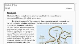
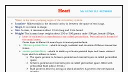
For B.Sc 4th Sem, ZOOH, , Genome, , Fish Heart:, The heart of Labeo is single circuit type. It always filled with venous blood or, deoxygenated blood, so it is called venous heart., The heart is composed of four chamber a sinus venosus an auricle a ventricle and, the fourth chamber is bulbus arteriosus. The heart receives the blood from the whole, body through the ductus cuvieri into, the sinus venosus. The sinus venosus is, proportionately larger and bears a pair, of lateral appendages, it is spongy in, nature. The sinus venosus is connected, with the auricle through a valve known, as sino-atrial valve. The auricle is, connected with the ventricle through the, auriculo-ventricular valve. The, ventricle is thick walled and muscular,, the ventricle supply blood to the gill via, bulbus arteriosus then through the, ventral aorta. The ventricle and the, bulbus arteriosus are connected each, other by a valve known as ventriculo-bulbar valve. The bulbus arteriosus is non, contractile, thin walled with smooth muscle and elastic fibers, but it lacks both cardiac, muscle and conal valves.
Page 2 :
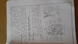
Amphibian Heart:, Structure:, The heart is the central pumping station for the circulation of blood. It is a hollow, pearshaped, muscular organ which is situated in the anterior part of the body cavity, in front of, the liver. It is completely enclosed in a transparent bag of membrane, the pericardium. The, broad base of the heart is directed forwards, whereas the narrow apex points towards the, posterior end and lies between the two main lobes of the liver., The heart is mainly composed of three chambers: a thick-walled, conical ventricle and two, thin-walled auricles, right and left. There are two other smaller chambers: a thin-walled, triangular sinus venosus on the dorsal side, opening into the right auricle, and a thick-walled, tubular conus arteriosus ventrally, connected to the base of the ventricle., The sinus venosus is a thin-walled sac-like chamber which is situated on the dorsal side of, the heart. It is more or less triangular in shape with the three caval veins opening into its, three corners. These veins carry deoxygenated blood into the sinus., It communicates with the right auricle through a slit-like sinu-auricular aperture, the edges, of which are guarded by the sinu-auricular valves. The valves permit entry of blood from, the sinus into the right auricle, but no backflow or regurgitation is allowed., The two auricles, right and left, form the base of the heart. They are separated from the, ventricle by a narrow groove, the coronary sulcus. The auricles are completely separated, from one another by a strong vertical partition, the inter auricular septum. The right auricle, is larger than the left; it receives deoxygenated blood from the sinus venosus through the, sinuauricular aperture., The left auricle receives oxygenated blood from the lungs through a small opening, the, aperture of the common pulmonary vein. The two auricles communicate with the ventricle, by a common opening, the auriculo-ventricular aperture, which is guarded by the auriculoventricular valves., The membranous cusps of this valve hang down like curtains to allow the passage of blood, from the auricles into the ventricle, but not in a reverse direction. The free edges of the, cusps are attached to the ventricular wall by means of a number of fine threads called, chordae tendineae., , The ventricle is the thick-walled conical chamber which is situated behind the auricles. Its, posterior bluntly pointed portion forms the apex of the heart. The ventricular cavity is, greatly reduced by a number of interlacing muscle fibres which arise from its own wall., The ventricle, therefore, is spongy and when it is cut, its interior looks like a honeycomb., The sponginess of the ventricle prevents admixture of the two kinds of blood in the, ventricular cavity, the oxygenated kind from the left auricle and the deoxygenated kind, from the right auricle., The conus arteriosus is the stout tube which arises ventrally from the base of the ventricle, and passes obliquely towards the left. It is continued forwards as the truncus arteriosus, which is the base of the main artery for carrying the blood away. The truncus however is
Page 3 :
not a part of the heart; it belongs to the arterial system. The conus is separated from the, truncus by a set of pocket-like semilunar valves., A similar set of three semilunar valves guards the opening between the ventricle and the, conus. These valves prevent backflow into the ventricle. Inside the conus is a twisted,, longitudinal flap, the spiral valve. This divides the cavity of the conus into a right channel,, the cavum aorticum, and a left channel, the cavum pulmocutaneum., The spiral valve, the semilunar valves and the spongy ventricular cavity co-operate with, one another in guiding the two kinds of blood. The deoxygenated blood is pumped through, the cavum pulmocutaneum and the oxygenated kind through the cavum aorticum. The two, kinds of blood enter different arterial arches and are carried to different places., , : Heart of Toad :, , : Inner structure and circulation through Heart :
Page 4 :
Structure of Reptilian Heart:, The heart of Calotes lies in the pleuroperitoneal cavity and occupies a position which is, midway between the fore-limbs though the ventricle extends slightly beyond the level of, the axillae. Thus the position of the heart is rather forward and such a disposition indicates, a lower grade of organisation because such condition is observed in Sphenodon., The heart is covered by a thin and transparent pericardial membrane. The space between, the heart and pericardium is filled with pericardial fluid. The heart is triangular in shape., The auri-cular region is wider than the ventricular region., Sinus venosus:, The heart is made up of five chambers. The sinus venosus is reduced and is disposed, transversely and dorsal to the lower half of the auricles. It is thin-walled and is formed by, the confluence of the venae cavae. The right half of the sinus venosus is larger than its left, counterpart and is formed by the confluence of right anterior vena cava (precaval vein) and, posterior vena cava (post-caval vein)., The left portion of the sinus venosus is composed mainly by the left anterior vena cava. A, constriction marks off the right and left parts of sinus venosus. The sinus venosus opens, into the right auricle near the region of the constriction by a semicircular sinuauricular, aperture., The aperture is provided with sinuauricular valves. The valves develop from the upper and, lower margins of the aperture and the free end of the valves which is slightly frilled projects, into the lumen of the right auricle., Auricle:, The right auricle is larger than the left auricle and appears darker than the left auricle. The, wall of the right auricle is thick and its inner lining is thrown into a number of musculi, pectinati which are projected within the lumen., The left auricle is smaller than the right auricle and it receives a common pulmonary vein., The pulmonary aperture is circular in outline and is located on its dorsal wall close to the, inter-auricular septum. The aperture is not provided with valves., Internally the left and right auricles are separated by a thin, muscular and non-perforated, inter-auricular septum. The septum extends posteriorly for a short distance within the, ventricle and bears at its posterior tip the auriculoventricular valves., Ventricle:, The ventricle is muscular, spongy and triangular in appearance. Its apex is directed caudal, and bears a thin and white cord of tissue, called gubernaculum cordis. It penetrates the, pericardium and reaches the upper margin of liver., The thick-walled ventricle is provided internally with an inter-ventricular septum which, divides it incompletely into left and right halves.
Page 5 :
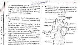
*This partition has become complete in crocodiles except for an aperture, called foramen, of Panizza. The foramen of Panizza is a communicating aperture between the left and right, systemic arches just at the point of crossing after their emergence from the ventricle., The inner cavity of the ventricle has been arbitrarily divided into three regions, namely,, cavum pulmonale situated in the right side, cavum arteriosum (or the left hand portion), by a muscular ridge and cavum venosum. The ridge arises from the right ventral wall of, the ventricle and runs dorsal obliquely. Higher up, it becomes horizontally inclined., It then runs obliquely again and takes a vertical course. It cuts off the aforesaid cavities., The left and right auriculoventricular apertures lie very close together being separated by, the prolongation of the inter-auricular septum. The free lateral edge of the septum bears the, auriculoventricular valves., In the middle of the ventricle and close to the line of demarcation between the auricles and, ventricle, there are three apertures from which the aortic arches arise. The inner wall of the, ventricle is provided with thick interlacing muscles, called columnae carnae. There are, bunches of thread-like muscle fibres, called chordae tendineae, by which the valves remain, attached to the columnae carnae., Valve:, The different compartments of the heart are intercommunicated by apertures having swing, door-like flaps, called the valves. These valves control the passage of blood and direct the, flow in one direction., The valves present in the heart of Calotes are:, (a) A pair of leaf-like valves in the sinuauricular aperture., (b) Sphincter muscle acting as valve in the opening of pulmonary vein into the left auricle,, (c) A pair of leaf-like valves formed by the bifurcation of the inter-auricular septum in the, auriculoventricular aperture,, (d) Three semilunar valves in each orifice from which the arterial arches arise., The walls of the heart are provided with three histological layers common to all blood, vessels,, i.e., tunica intima, tunica media and tunica adventitia from inner to the outside. Of, these three layers, the tunica media is peculiar in having specialised cardiac muscles, showing striations and branching’s. The heart is supplied with the cardiac branch of 10th, cranial nerve., , Mechanism of circulation through heart:, In Calotes, the circulatory circuit is double. There are pulmonary or lesser circulation and, systemic or greater circulation. Pulmonary circulation is conducted by the pulmonary, arteries which carry deoxygenated blood to the lungs. In the lungs, the blood becomes, oxygenated and returns to the left auricle by the pulmonary vein.
Page 6 :
The left auricle pours its content into the ventricle through the auriculoventricular aperture., In the greater circulation, deoxygenated blood returns to the sinus venosus by two precaval, and one postcaval veins. The sinus venosus opens into the right auricle. The right auricle, empties its content into the ventricle., The ventricle sends blood for circulation into the different parts of the body through the, systemic and pulmonary arches. The entry and exit of blood in the ventricle are so, beautifully arranged that a major quantity of oxygenated blood is always forwarded to the, brain region., As the ventricle is incompletely divided, admixture of oxygenated and deoxygenated blood, occurs thrice in Calotes once in the cavum venosum, once in the dorsal aorta and another, in the left ductus caroticus., Thus, though the ventricle in Calotes is morphologically incompletely divided, there is a, tendency for the physiological separation of the two types of blood, at least in two, auricles com-pletely and in the ventricle partially. From this point, the heart of Calotes is, biologically more advanced than that of Bufo., , Fig: Heart of Squamata (Calotes), , Fig: Crocodilian Heart